
Vascular Conditions
All Vascular Conditions
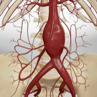
Abdominal Aortic Aneurysms
Vascular Conditions
What is Abdominal Aortic Aneurysms? Abdominal Aortic Aneurysms (AAA) are caused by progressive weakening of the aortic wall creating a “ballooning” of the vessel. While most aneurysms are asymptomatic and do not cause problems, some may expand in size and eventually rupture if not diagnosed and treated. Aneurysms occur most often in the aorta, the main artery of the chest and abdomen. The aorta carries blood from the heart to all parts of the body including the vital organs and the legs and feet. Symptons In most cases, there are no major symptoms for AAA. Occasionally, patients may feel abdominal, back or side pain. 75% of the aneurysms that are discovered are detected from diagnostic tests (such as x-rays) that were given for other health problems. Individuals may also feel a coldness, numbness or tingling in their feet and hands. A ruptured aneurysm is an emergency and procedures must take place immediately to save one’s life and avoid serious complications. Diagnosis Although AAA can be detected by physical examination, most are diagnosed today using an ultrasound scan or CAT scan. These are simple, non-invasive exams conducted on an outpatient basis. These exams also measure the size of AAA, a key element in determining the best treatment. Early Diagnosis When aortic aneurysms are diagnosed early, treatment is safe and effective and the aneurysm is curable. Aneurysms are often detected while performing tests for entirely different reasons. Most patients have no symptoms, so if you are at risk, it is important to discuss AAA with your doctor. Risk Factors Age over 60 years Tobacco use Family history of AAA History of heart disease or peripheral artery disease High blood pressure (hypertension) Causes Proteins in the wall of the aorta, called elastin and collagen, provide strength and flexibility to this large artery. This is similar to muscles and tendons providing strength to the arms and legs. Aneurysms are caused by a progressive breakdown of these proteins that lead to a weakness of the wall of the aorta that can steadily expand like a balloon. These proteins, collagen and elastin, may gradually deteriorate with age. Inflammation that is associated with atherosclerosis (hardening of the arteries) helps to accelerate this degenerative process even in younger people. Some of the body’s naturally occurring enzymes may also cause the breakdown of collagen and elastin in the wall of the aorta. An excess of these enzymes or conditions that activate the enzymes may cause the formation of an aneurysm, or lead to its sudden growth. In rare cases an aneurysm may be caused by infection. There is still much to be learned about the cause of aneurysms and their growth. Treatment If the AAA is larger than 5-6 centimeters in diameter (about the size of a lemon), it will require treatment. Your physician’s decision to repair will be based on the risk of the aneurysm rupturing, along with surgical or procedure risks, and risks associated with other pre-existing conditions. Smaller aneurysms that cause back or abdominal pain may also need treatment, especially for those that are enlarging rapidly. Surgical treatment of AAA has been performed routinely in the U.S. for about 50 years. It is a very successful and durable procedure. During the surgery, the surgeon makes an abdominal incision, then replaces the diseased part of the aorta with a Dacron or Teflon graft that is carefully matched to the normal aorta. This graft is sewn in place by the surgeon. Most patients stay in the hospital for five to ten days if no complications occur. Complete recovery from the operation may take one to two months before returning to a full and normal life. After 50 years of experience with these procedures, the facts show that more than 90% of patients make a full recovery from surgery. Once patients have recovered, their aneurysms are permanently cured! Less Invasive Treatments of AAA Recent advances in catheter-based technologies have led to exciting new treatments for aortic aneurysms. Now, endovascular grafting technology allows the repair of the AAA by inserting a graft through a small incision in the groin. The endovascular method allows the graft to be delivered through a catheter or tube inserted in a groin artery. X-ray guidance is then used to accurately position the graft in the AAA. The graft is then expanded inside the aorta and held in place with metallic hooks rather than sutures. The hospital stay is usually only one or two days, and most patients can return to work or normal daily activities in about a week. Patients with other medical problems or those that could not withstand major surgery can be considered for repair by an endovascular graft. Endovascular grafting may not be possible in every case. Endovascular grafts are specially manufactured and don’t “fit” everyone’s anatomical situation. Standard surgery may still be the best option for many. As a fairly new procedure, endografts do not yet have a 50-year track record to compare to that of surgery for AAA. Speak with your physician about the best option for you. The combination of early diagnosis with safer, simpler, and ever more successful treatments can prevent deaths due to ruptured abdominal aortic aneurysms. Fast Facts • The risk of AAA increases with age • Tobacco users are eight times more likely to be affected than non-users • AAAs are between five to ten times more common in men than in women • There are approximately 15,000 deaths per year related to the rupture of an aneurysm • Ruptured aneurysms are the 10th leading cause of death in men over 50 in the country. Source: https://vascularcures.org/abdominal-aortic-aneurysm/ Credit: Vascular Cures

Abdominal Aortic Aneurysms VHC
SVS Vascular Conditions
Abdominal Aortic Aneurysms - What is an AAA? The aorta is the largest artery in your body. It starts at your heart and runs through your chest and abdomen. The abdominal area of the aorta is responsible for delivering blood to your legs, gastrointestinal tract, and kidneys. An abdominal aortic aneurysm (AAA) happens when the wall of the aorta weakens over time and begins to bulge like a balloon. If left untreated, an AAA may get bigger and eventually rupture, causing severe internal bleeding and possibly death. In addition to the risk of rupturing, clots or debris can form within an AAA. These pieces can be carried to other areas in the body and block circulation, which can cause severe pain or even limb loss if blood flow is cut off for too long. Every year, 200,000 people in the U.S. are diagnosed with an abdominal aortic aneurysm (AAA). A ruptured AAA is the 15th leading cause of death in the country, and the 10th leading cause of death in men older than 55. Aneurysms often run in families. If a first-degree relative, like a parent or sibling, has had an AAA, you are 12 times more likely to develop an AAA. About 15-25% of people who need treatment to repair an AAA have an immediate family member with the same type of aneurysm. Learn More Symptoms No Symptoms : Most people with an AAA have no symptoms and it is often found when tests are performed for unrelated reasons. Sudden, Severe Abdominal or Back Pain : If you have a family history of AAA and feel sudden, severe pain in your abdomen or back, seek immediate care. This may signal that you have an AAA and it is possibly rupturing. Pain, Discolored Skin, Sores on Feet and Toes : A small percentage of patients have these symptoms when plaque or blood clots from elsewhere in the body collect in the feet and toes. Causes and Risks Older Age Males are more at risk than females Family History of AAA Smoking High blood pressure (Hypertension) Atherosclerosis (hardening of the arteries) Chronic lung disease Talk to a Doctor If you have risk factors for an AAA or have been diagnosed with one, you should talk to your doctor about a referral to see a Vascular Surgeon. Diagnosis If an AAA is suspected, your doctor may refer you for: An abdominal ultrasound , which is a painless, safe test that screens for and measures the size of the AAA. A computed tomographic angiogram (CTA) might also be recommended, which assesses the aneurysm size, location and the extent of impact. It would expose you to radiation and injection of an intravenous contrast agent. However, it provides valuable information that can help your vascular surgeon determine the best type of repair. Role of a Vascular Surgeon If you’re diagnosed with an AAA, you should be referred to a vascular surgeon. They can determine when treatment is needed and recommend the best procedure for patients with AAA, based on the size of the aneurysm and other factors. Because vascular surgeons have specific training in both minimally invasive and traditional open repairs, they can help ensure that the repair is tailored to your unique anatomy and expectations. Treatments Treatment for AAA depends on the size of the aneurysm. Small AAAs (Less than 5 cm in diameter) Small AAAs have a very low risk of rupturing and should be watched. It’s important to have an ultrasound test every 6-12 months to check for growth and risk of rupture. Treatment can include: Lifestyle changes that help control blood pressure and medication Quitting smoking Daily exercise Larger AAAs (More than 5.0-5.5 cm in diameter) Larger, rapidly growing AAAs and ones causing symptoms are usually repaired. Types of repair include: Endovascular aneurysm repair (EVAR) Endovascular aneurysm repair (EVAR) is a less invasive treatment and is most frequently performed. Your vascular surgeon would make two small punctures in your groin. Guided by X-ray imaging, a tiny device is introduced into the artery. This device is used to reinforce the artery wall and exclude the aneurysm. Most patients stay in the hospital one to two days. Recovery time is shorter than with open surgery. Open Surgery Open surgery is performed to place a prosthetic graft. Your vascular surgeon would access the affected portion of the aortic artery through an incision in your abdomen. Most patients stay in the hospital four to 10 days. Recovery time may take up to three months. Source: https://vascular.org/your-vascular-health/vascular-conditions/common-conditions/abdominal-aortic-aneurysm
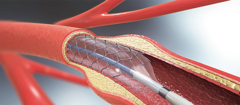
Acute Limb Ischemia
SVS Vascular Conditions
Acute Limb Ischemia Acute limb ischemia (ALI) occurs when there is a sudden lack of blood flow to a limb, typically an arm or leg. This can be due to a blood clot or a build-up of fatty deposits inside the arteries, causing them to narrow —like what occurs in heart disease. Without prompt treatment, this condition can lead to tissue damage, organ dysfunction, and loss of the affected limb. ALI is a medical emergency and requires immediate care. Symptoms ALI symptoms are often severe and sudden. They can be remembered with the "six Ps": Pain : sudden severe pain in the limb, even when at rest Pallor : the limb may appear pale Pulselessness : absent or diminished pulse in the limb Paresthesia : a tingling or prickling sensation, or numbness Paralysis : loss of movement in the limb, which is a late and grave sign Poikilothermia : the limb feels colder than the rest of the body Causes and Risks ALI can be caused by the following: Embolism : A blood clot from elsewhere in the body blocks a limb artery. Thrombosis : A clot forms inside an artery that is already narrowed by plaque. Trauma : Injury to an artery. ALI risk factors include heart conditions, irregular heartbeat, atherosclerosis, previous clots, and smoking. Diagnosis - Talking to a Doctor If a patient experiences any of the previously described symptoms, they should seek medical attention immediately. ALI is an emergency condition. Patients should discuss any risk factors with their doctors and seek a referral to a vascular surgeon if concerned about ALI. Role of Vascular Surgeon Vascular surgeons are experts in diagnosing and treating vascular conditions such as ALI. Vascular surgeons are trained to perform emergency procedures that can restore blood flow and save the affected limb. Diagnosis Diagnosis usually involves urgent assessment by a vascular surgeon and may include: During physical examination, checking for the presence of pulses in the limb. Conducting Doppler ultrasound to assess blood flow and locate any blockages. Angiography, which involves injecting a contrast dye into the arteries to visualize blockages on X-ray images. Using CT scan to identify blockages of the blood vessels. Treatments Treatment aims to restore blood flow quickly. It may include: Medications to dissolve clots or prevent new ones Catheter-directed thrombolysis to deliver clot-dissolving drugs directly to the site of the clot Catheter-based thrombectomy, which uses a tube inserted into the artery to remove the blood clot Surgical procedures to remove the clot or bypass the blocked artery Angioplasty or stenting to open the narrowed arteries and maintain blood flow Time is critical in treating ALI to prevent permanent damage. If a patient suspects they are experiencing ALI, they should seek immediate emergency care. Source: https://vascular.org/patients-and-referring-physicians/conditions/acute-limb-ischemia
Aortic Dissection
Vascular Conditions
What is Aortic Dissection (AD)? Aortic dissection is the most common catastrophe affecting the aorta. The aorta is the largest artery of the body through which blood leaves the heart to deliver oxygenated blood to the rest of the body. It occurs in about 24 people per million each year in the U.S. It is caused when the inner layer of the aortic wall tears and then peels or separates away from the next layer of the aorta. This creates two channels; the original aortic channel for blood flow (the true lumen) is still present while the peeling away of the outer layer in the dissection creates a new additional ow channel (the false lumen). This double-barrel flow pattern in the dissected aorta can cause serious problems upstream or downstream from the tear. The dissection or separation can result in a significant decrease in blood flow to various organs and tissues supplied by the involved branches. Branches that may be affected include the kidney (renal) arteries, the gut (mesenteric) arteries, the arteries to the brain, and the arteries to the arms or legs. In addition, the tear may compromise blood flow to the heart causing a heart attack and in some cases can result in internal bleeding around the heart, causing loss of consciousness or death. Aortic dissection is a very serious, life-threatening disorder that requires immediate medical attention. Risk Factors The main preventable risk factor for an aortic dissection is high blood pressure (hypertension). Over time, hypertension weakens the aortic wall, making it more likely to have a spontaneous tear involving the inner lining of the aorta. The tear exposes the middle of the wall to very high aortic blood pressure. This creates a separation that unzips the aorta between the layers which are pulled apart by the dissection process, leading to the double-barrel flow pattern. Other risk factors for aortic dissection include: Marfan or Ehlers-Danlos syndrome, which are inherited disorders of blood vessels and connective tissues; presence of aortic aneurysm or enlargement of the aorta; a family history of aortic dissection; and certain heart conditions such as a bicuspid aortic valve. Aortic dissection may also occur as a complication of open heart surgery or heart catheterization, although this is very rare. Symptons Location of Pain Chest pain Back pain Flank pain Abdominal pain Leg pain Quality of Pain Pain that is tearing or sharp Abrupt onset of pain Pain that migrates or radiates Neurological Deficits Coma/Altered consciousness Paralysis Extremity numbness Difficulty with speaking or slurred speech Light-headedness/Fainting Difficulty breathing/Shortness of breath Diagnosis When a physician suspects the diagnosis of aortic dissection, an imaging test is generally ordered to evaluate the aorta. There are three primary tests used to diagnose aortic dissection. Aortic dissection can be diagnosed with a CAT scan of the aorta or an MRI scan. A transesophageal echo (TEE) may also be performed. A TEE is a special type of ultrasound test during which a small ultrasound probe is passed through the mouth and into the stomach and esophagus to take very detailed pictures of the heart and aorta. Currently there are no blood tests that can accurately diagnose aortic dissection. Treatment In all cases, the first treatment for patients with aortic dissection is aggressive control of blood pressure with medications, usually given through an intravenous line (IV). Patients with aortic dissection are generally monitored in the intensive care unit. A team of experts cares for patients with aortic dissection, including emergency medicine physicians, cardiac and vascular surgeons, cardiologists, radiologists, and internal medicine specialists. The treatment of aortic dissection depends upon a number of factors, including the location of the tear in the aortic wall, how much of the aorta is involved, the effects of the dissection on the branch vessels of the aorta and the perfusion of the organs, and the patient’s symptoms. For patients with aortic dissection who require invasive treatment, surgery to repair the dissected portion of the aorta is generally the recommended treatment. In some cases, minimally invasive stenting procedures can be used to treat aortic dissection. For all patients who have an aortic dissection long-term follow-up with a physician is a very important part of treatment. Blood pressure and heart rate need to be carefully monitored and controlled with medications. For many patients, CAT scans or MRI scans will be repeated at regular intervals (such as every 6 months or every year) to monitor the size of the aorta and the status of the dissection. Source: https://vascularcures.org/vascular_diseases/aortic-dissection-ad/ Credit: Vascular Cures

Aortic Dissection VHC
SVS Vascular Conditions
Aortic Dissection Aortic dissection is a severe condition in which a tear develops in the inner layer of the body's main artery, the aorta. This tear causes blood to rush through, splitting the aorta’s inner and middle layers. If the blood breaches the outer aortic wall, it is usually fatal. Learn More While this disease is rare, it can be seen in men who are in their 60s and 70s. Aortic dissection is classified into two types based on the location of the tear. Type A dissections , which are more common and severe, involve a tear in the aorta near the heart or in the upper aorta, known as the ascending aorta. This type of dissection can potentially extend into the abdomen. Type B dissections involve a tear in the lower portion of the aorta, known as the descending aorta. These can also extend into the abdomen. Both types of aortic dissection pose significant risks, including the potential for life-threatening complications if not promptly treated. Symptoms can mimic other illnesses, leading to delayed diagnosis. Early detection and treatment significantly increase survival chances. Symptoms Symptoms of aortic dissection can resemble those of other heart issues, such as a heart attack. Common signs include: Sudden, intense chest or upper back pain, often described as tearing or ripping, which spreads to the neck or back Acute stomach pain Fainting Breathlessness Stroke-like symptoms, such as sudden vision problems, speech difficulty, or one-sided body weakness Weaker pulse in one arm or thigh compared to the other Leg discomfort or weakness Walking difficulty Causes and Risks High blood pressure, cocaine use, smoking and some genetic connective tissue disorders make dissections more likely to occur. Talking to a Doctor Patients should discuss with their doctor any of the concerning symptoms listed previously and ask if referral to a vascular surgeon should be done. Early detection and prompt treatment are crucial for aortic dissection, so patients should seek medical attention as soon as symptoms arise. See a Vascular Surgeon For patients suffering from aortic dissections, vascular surgeons provide timely and appropriate diagnosis. They are educated and trained in endovascular and surgical intervention to repair the tear in the aorta. This may involve open surgery or stents to repair the damaged artery and prevent further complications. Additionally, the vascular surgeon collaborates closely with other members of the healthcare team to ensure comprehensive care for the patient, including preoperative assessment, postoperative management, and long-term follow-up to monitor for any potential complications. Diagnosis Diagnosing an aortic dissection typically involves a combination of medical history, physical examination, and diagnostic tests. Physicians often begin by assessing the patient's symptoms and medical history, paying close attention to any sudden or severe chest or back pain. A physical examination may reveal indications, such as differences in blood pressure between the arms or unequal pulses in the limbs. To confirm the diagnosis and determine the extent of the dissection, various imaging tests are used, including computed tomography (CT) scans, magnetic resonance imaging (MRI), or echocardiography . These tests help visualize the structure of the aorta and identify any tears or abnormalities. Early and accurate diagnosis is crucial for initiating appropriate treatment and improving patient outcomes. Treatments Treatment for aortic dissection aims to stabilize the patient's condition, relieve symptoms, prevent complications, and repair the damaged aorta, if possible. In most cases, emergency surgery is necessary, particularly for Type A dissections involving the ascending aorta. The surgical procedure typically involves replacing the damaged portion of the aorta with a synthetic graft to restore normal blood flow. For Type B dissections, the approach may involve medications to control blood pressure and heart rate, in addition to close monitoring to prevent further complications. In some cases, endovascular repair procedures, such as stent grafting, may be considered as an alternative to open surgery. The choice of treatment depends on various factors, including the extent and location of the dissection, the patient's overall health, and the presence of any complications. Prompt medical intervention is essential to improve the prognosis and prevent life-threatening complications associated with aortic dissection. Staying Healthy - To minimize the risk of an aortic dissection: Have your blood pressure tested regularly. If you have high blood pressure, follow your doctor’s instructions for managing it. Avoid use of recreational drugs. If you don’t smoke, don’t start. If you do smoke, ask your vascular surgeon to help you find a smoking cessation program that will work for you. Source: https://vascular.org/your-vascular-health/vascular-conditions/aortic-dissection
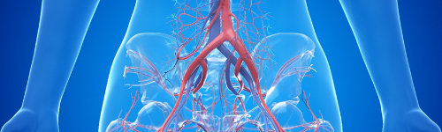
Aortoiliac Occlusive Disease
SVS Vascular Conditions
Aortoiliac Occlusive Disease ortoiliac occlusive disease is the narrowing or blockage of the aorta, the main blood vessel in your body, or the iliac arteries. The iliac arteries are the branches that your aorta divides into around the level of the belly button to provide blood to your legs and the organs in your pelvis. This blockage is typically caused by a buildup of plaque within the walls of your blood vessels. The aorta and iliac arteries are the second most common blood vessels to be affected by peripheral arterial disease (PAD) after the blood vessels in the thigh. PAD occurs in 12-20% of people over the age of 65. Illiac artery is narrowed by plaque. Causes and Risks The most common cause of aortoiliac disease is atherosclerosis (hardening of the arteries). Hardening of the arteries may be caused by smoking, high cholesterol, high blood pressure, diabetes, genetic predisposition or obesity. Inflammatory conditions such as Takayasu’s arteritis may cause inflammation in the arteries causing blockages of the arteries. Radiation to the pelvis may cause progressive inflammation in the arterial wall leading to blockages of the arteries. Symptoms May Be Absent You may have varied symptoms or no symptoms at all and still have aortoiliac occlusive disease. Fatigue, Pain, or Cramping When Walking When felt in the buttocks, thighs or calves, these symptoms may signal mild to moderate aortoiliac occlusive disease. As the disease progresses, these symptoms may occur after walking very short distances. Erectile Dysfunction Men may experience erectile dysfunction. Pain at Rest, Leg and Feet Problems In severe cases, you may feel pain in your feet or toes at rest, coldness and numbness in your legs, sores or wounds on your legs and feet, and even gangrene or the death of tissue in your feet. Diagnosis - See a Vascular Surgeon You will be asked questions about symptoms and medical history, including questions about family members. The vascular surgeon will also perform a physical exam. Tests May Be Recommended Initially an ankle-brachial index test and a duplex ultrasound may be recommended. If further testing is needed a CT angiogram, MR angiogram, or a catheter-directed angiogram are options. These studies use different types of dye to help identify where blockages in the arteries are located to plan further treatment. With a CT angiogram and MR angiogram, dye is delivered through an IV. With a catheter-directed angiogram, dye is injected through a thin tube (catheter) inserted in an artery. Treatments Aortoiliac occlusive disease may be managed by: Risk factor modification, such as quitting smoking, controlling cholesterol or high blood pressure, managing diabetes and regular exercise. Medication may be prescribed, such as an aspirin or another medication to prevent platelets from clotting in the blood. A statin drug helps to control cholesterol and help prevent plaque progression. If further treatment is needed, a minimally invasive procedure or a surgical bypass may be considered. The most common minimally invasive treatment is the placement of a stent in your aorta or iliac arteries. This procedure is commonly done at the same time as a catheter-directed angiogram. The stent is a small device that opens the blockage in the arteries to create a wider path for blood flow to the lower half of your body. An angioplasty, in which an inflatable balloon device is inserted through the catheter to further assist in opening up the arteries, can be added to the treatment. A surgical bypass is the creation of a detour around the blockage. The detour may start in the aorta and may end either in the iliac arteries in your pelvis, or in an artery in your groin or leg. A bypass may be performed either on one or both sides at the same time, usually using an artificial graft. If there is not a suitable artery to originate the bypass in the aorta, or if you are not healthy enough to undergo an aortic-based procedure, the bypass generally starts at the axillary artery underneath the collarbone and ends in one or both legs. Staying Healthy The best way to prevent development of aortoiliac occlusive disease is to not smoke, and manage cholesterol, blood pressure and diabetes. If you do smoke, ask your vascular surgeon to help you find a smoking cessation program that will work for you. Get regular exercise. Eat a healthy, well-balanced diet that is low in fat and cholesterol. Source: https://vascular.org/your-vascular-health/vascular-conditions/aortoiliac-occlusive-disease
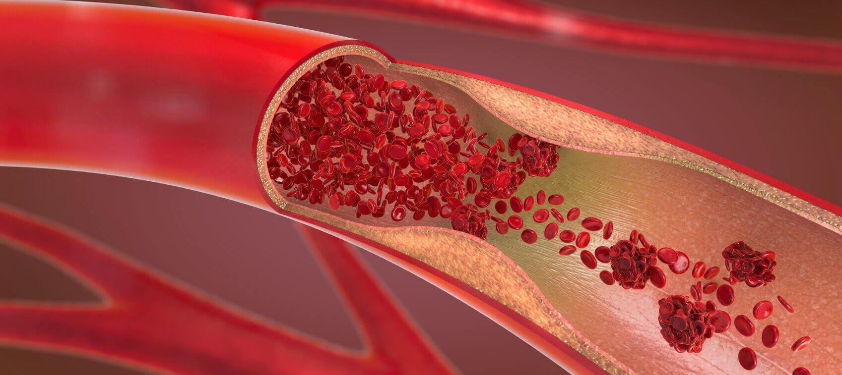
Arm Artery Disease
SVS Vascular Conditions
Arm Artery Disease Arm artery disease is rare, and usually indicates other health issues. Typically, blockages in your arm arteries occur when blood clots travel there from your heart or from an injured artery within your chest. Symptoms Severe Pain and Numbness in the Hands These symptoms may indicate a fresh blood clot in the arm, which is a medical emergency. Call 911 if you have these symptoms. Painful or Numb Hands or Fingers Coldness and numbness followed by tingling or stinging pain in your fingers may signal Raynaud’s Disease that is linked to a disorder in your arm arteries. A dialysis access fistula or graft may take blood away from your hand (steal syndrome), resulting in hand pain or numbness. Forearm Cramping or Sores That Won’t Heal These are additional symptoms of arterial disease in the arm. Causes and Risks Arterial disease of the arm can be caused by atherosclerosis , autoimmune diseases, complications from dialysis access or by blood clots that travels from the heart (embolism). Diagnosis - See a Vascular Surgeon You will be asked questions about symptoms and medical history, including questions about family members. The vascular surgeon will also perform a physical exam that will likely include taking blood pressure readings in your arms and in your fingers. Tests May Be Recommended A duplex ultrasound can aid diagnosis. If more information is needed, a CT scan or angiogram may be recommended. Treatments Treatment Depends on the Source of Symptoms Atherosclerotic blockages can be treated with angioplasty, stenting or surgical bypass . Autoimmune conditions are usually treated with medicines. A fresh clot in the arm is treated with lysis ( thrombolytic therapy ) to break up blood clots, or with open surgery to extract the clots. If the lack of blood flow is due to dialysis access , there are a variety of procedures to increase hand blood flow and possibly preserve the fistula or graft. Staying Healthy Avoiding smoking. If you do smoke, ask your vascular surgeon to help you find a smoking cessation program that will work for you. Keep your cholesterol down. Maintain a healthy weight. If you have a heart arrhythmia like atrial fibrillation, it is important to take blood thinners if prescribed by your doctor. Source: https://vascular.org/your-vascular-health/vascular-conditions/arm-artery-disease

Arterial Dissection VHC
SVS Vascular Conditions
Arterial Dissection Arterial dissection occurs when a tear forms in the innermost layer of an artery, allowing blood to enter and separate the layers of the arterial wall. This condition can affect arteries throughout the body, including the carotid arteries in the neck and the arteries supplying the brain. Arterial dissection causes a disruption in blood flow and can potentially lead to serious complications, such as stroke or organ damage. Arterial dissection can be spontaneous or triggered by trauma, connective tissue disorders , or certain activities. Early diagnosis and intervention are crucial for preventing complications and improving outcomes in patients with arterial dissection. Learn More Symptoms Symptoms of arterial dissection can vary depending on the location and severity of the tear in the artery. Common symptoms include: sudden, severe headache neck pain or stiffness facial droop or asymmetry vision changes or loss of vision in one eye weakness, numbness, or tingling on one side of the body difficulty speaking or understanding speech dizziness or loss of balance hearing a rhythmic sound in the ears (pulsatile tinnitus) fainting or loss of consciousness chest or back pain sudden onset of weakness in the legs Arterial dissection symptoms can develop rapidly and may worsen over time, particularly if the dissection leads to reduced blood flow to vital organs. If any of these symptoms occur suddenly or are severe, patients should seek immediate medical attention. Causes and Risks While the exact cause of arterial dissection is not always clear, several factors and conditions may increase the risk, including: Trauma : Injury or trauma to the artery, such as from a car accident, sports injury, or blunt force trauma, can cause arterial dissection. Connective tissue disorders : Certain connective tissue disorders, such as Ehlers-Danlos syndrome, Marfan syndrome, or fibromuscular dysplasia, can weaken the arterial walls and predispose them to dissection. High blood pressure : Chronic hypertension can put increased pressure on the arterial walls, making them more susceptible to tearing. Atherosclerosis : The buildup of plaque ( atherosclerosis ) within the arteries can lead to weakening and damage to the arterial walls, increasing the risk of dissection. Genetic factors : There may be a genetic predisposition to arterial dissection if individuals have a family history of the condition. Pregnancy and childbirth : The hormonal changes and physical stress associated with pregnancy and childbirth may increase the risk of arterial dissection in some women. Certain medical procedures : In rare cases, medical procedures such as angiography, angioplasty, or chiropractic manipulation may trigger arterial dissection. While arterial dissection can occur spontaneously, certain risk factors may increase the likelihood of its development. It's important for patients to be aware of these risk factors and seek medical attention immediately if they experience symptoms. Talking to a Doctor Discuss symptoms of arterial dissection with a doctor as soon as possible, especially if symptoms are sudden, severe, or persistent. Patients should seek immediate medical attention if they experience any of the symptoms previously described. Diagnosis Early recognition and treatment of arterial dissection can help prevent complications and improve outcomes. Diagnosing arterial dissection typically involves a combination of clinical evaluation, imaging studies, and diagnostic tests. The process may include the following steps: Medical history and physical examination Imaging studies: Imaging tests are essential for visualizing the structure and function of the affected artery. Common imaging modalities used in the diagnosis of arterial dissection include: Magnetic resonance imaging (MRI) and magnetic resonance angiography (MRA) Computed tomography angiography (CTA) Doppler ultrasound Catheter angiography Laboratory tests: Blood tests may be conducted to assess for signs of inflammation, coagulation disorders, or other underlying conditions that may contribute to arterial dissection. Once a diagnosis of arterial dissection is confirmed, further evaluation may be conducted to assess the extent of the dissection, determine the underlying cause, and guide treatment decisions. Prompt and accurate diagnosis is essential for initiating appropriate management and preventing complications associated with arterial dissection. Role of a Vascular Surgeon Overall, vascular surgeons play a pivotal role in the comprehensive management of arterial dissection. They offer expertise in diagnosis; treatment planning; and interventions aimed at restoring blood flow, preventing complications, and improving patient outcomes. Treatments The treatment of arterial dissection aims to relieve symptoms, prevent complications, and restore normal blood flow to the affected artery. Treatment is individualized based on the specific needs and circumstances of each patient and depends on several factors, including the location and severity of the dissection, the presence of complications, and the patient’s overall health. In many cases, medications are prescribed to manage symptoms and reduce the risk of complications. For example, blood pressure medications control hypertension and reduce stress on the arterial walls. Treatment might also include minimally invasive procedures to repair the arterial dissection and restore normal blood flow. These procedures include: Stent placement, when a small mesh tube is inserted into the affected artery to support the arterial wall and keep it open, allowing blood to flow freely. Balloon angioplasty, when a balloon catheter is used to widen the narrowed or blocked portion of the artery, improving blood flow. Thrombectomy or Thrombolysis, which is used in cases where a blood clot is causing or complicating the dissection. It involves using a catheter to remove the blood clot or thrombolytic medications administered directly into the artery to dissolve the clot. In some situations, open surgical repair may be necessary, particularly if the dissection is extensive or if endovascular therapy is not feasible. Surgical options include: Arterial bypass grafting, in which the surgeon creates a bypass to redirect blood flow around the affected artery, bypassing the dissection site. Arterial reconstruction, in which the surgeon removes the damaged portion of the artery and connects the remaining ends to restore blood flow. After treatment, patients with arterial dissection require close monitoring to assess the effectiveness of treatment, monitor for complications, and adjust medications as needed. Follow-up appointments with healthcare providers and imaging studies may be scheduled to evaluate the healing process and ensure long-term vascular health. Source: https://vascular.org/your-vascular-health/vascular-conditions/arterial-dissection
Arteriovenous Malformation
Vascular Conditions
What is Arteriovenous Malformation? Arteriovenous malformation (AVM) is a vascular disorder in which arteries and veins are connected directly rather than through capillaries. This direct connection produces enlarged, tangled masses creating a fast flow lesion within the blood vessels that is prone to rupture, bleeding, and stroke. AVMs can develop in many different areas of the body, including the lungs, kidney, liver, iris, spermatic cord and the areas between the ribs. 50% of AVMs occur in the brain or spinal cord can have especially widespread effects on the body. AVMs of the brain or spinal cord are believed to affect approximately 300,000 Americans. They occur in males and females of all racial or ethnic backgrounds at roughly equal rates. Causes Scientists believe that arteriovenous malformations are most often the result from mistakes that occur during embryonic or fetal development (congenital arteriovenous malformation). The genetic transmission patterns of AVM, if any, are unknown but often occur in multiples and have a female predominance. An estimated 300,000 Americans have AVMs, of whom 12% (approximately 36,000) will exhibit symptoms of greatly varying severity. Warning Signs Approximately 88% of people affected with AVM do not have any symptoms. The most general symptoms of a cerebral AVM include headache and seizures. More specific symptoms depend on the location of the malformation and the individual. These symptoms include: • Difficulties with movement or coordination, including muscle weakness and even paralysis • Vertigo (dizziness) • Difficulties with speech and communication • Difficulties with everyday activities • Abnormal sensations (numbness, tingling, or spontaneous pain) • Memory and thought-related problems, such as confusion, dementia or hallucinations The greatest potential danger posed by an AVM is hemorrhage. Researchers believe that each year between 2 and 4% of all AVMs hemorrhage. Treatment & Prevention Treatments for arteriovenous malformations vary on a case-by-case basis and depend on the size and location of the AVM. Treatments include the following: Medications can often alleviate general symptoms such as headache, back pain, and seizures, but are not a definitive treatment for an AVM. Conventional surgery involves removing the central portion of the AVM while causing as little damage as possible to surrounding structures. This surgery is most appropriate when the AVM is superficially located and relatively small in size. Radiosurgery is a less invasive therapeutic approach involving aiming a beam of highly focused radiation at the AVM. The radiation damages the walls of the blood vessels, creating a lesion. Over the course of the next several months, the irradiated vessels gradually degenerate and eventually close, leading to the resolution of the AVM. Endovascular embolization is less invasive than conventional surgery and involves the surgeon guiding a catheter through the arterial network until the tip reaches the site of the AVM. The surgeon then introduces a substance that will become lodged in a vessel of the AVM and obstruct blood flow. Since embolization does not permanently obliterate the AVM, it is usually used as an adjunct to surgery or radiosurgery to reduce the blood flow through the AVM and make surgery safer. Because so many variables are involved in treating AVMs, doctors must assess the danger posed to individuals on a case-by-case basis. The consquences of hemorrhage are potentially dangerous, leading many clinicians to recommend surgical interventon whenever the physical characteristics of an AVM appear to indicate the likelihood of significant bleeding and possible neurological damage. In some cases, it might be better to avoid treatment and simply accept a small risk of coming to harm from the AVM. This question is currently being addressed in clinical trials. Source: https://vascularcures.org/vascular_diseases/artiovenous-malformation/ Credit: Vascular Cures

Arteriovenous Malformation VHC
SVS Vascular Conditions
Arteriovenous Malformation Arteriovenous malformations (AVMs) are a complex tangle of abnormal arteries and veins that can occur anywhere in the body. Normally, arteries carry oxygen-rich blood from the heart to the brain, and veins carry oxygen-depleted blood back to the lungs and heart. But in an AVM, the arteries connect directly to veins via a tangle of blood vessels, disrupting this vital cycle. AVM can lead to various health issues, including bleeding, seizures, or other neurological symptoms. AVMs are congenital, meaning they are present at birth. Symptoms Many individuals with AVMs may not experience any symptoms. Depending on the location and size of the AVM, patients may experience pain or swelling. If the AVM involves the skin, it can ulcerate and bleed. Causes and Risks The exact cause of AVMs is unknown, but they are believed to form during fetal development. Certain genetic factors may play a role. Risks associated with AVMs include bleeding, seizures, and neurological deficits. The risk of rupture varies depending on the AVM. Talk to a Doctor If you experience or have concerns about AVMs, discuss them with your doctor. A referral to a specialist might be necessary for further evaluation. Role of a Vascular Surgeon Vascular surgeons are specialists that diagnose and treat AVMs. They collaborate with other specialists to determine the best approach to treat a patient’s condition. Diagnosis Diagnostic tests for an AVM might include vascular ultrasound, MRI or CT scans, or angiography. Treatments Treatment options depend on the location, size, and symptoms caused by the AVM, as well as the patient’s overall health. Treatment might include Medical management, which addresses AVM symptoms. Surgery, to remove the AVM if it is accessible and the risk of surgery is acceptable. Embolization, which is a minimally invasive endovascular procedure that reduces the size of the AVM or closes off the abnormal blood vessels. Stereotactic radiosurgery, a focused radiation treatment that aims to damage the vessels in the AVM and cause them to close off over time. Careful monitoring and follow-up care are critical for all patients with AVMs, whether they undergo treatment or not. Learn More Source: https://vascular.org/your-vascular-health/vascular-conditions/arteriovenous-malformations
Atherosclerosis Statistic Normal
Vascular Conditions
Atherosclerosis Statistic Normal vs. Diseased Arteries Atherosclerosis is a vascular disease that is caused by a buildup of plaque in the inner lining (intima) of arteries that restricts or blocks blood flow to a specific organ or region of the body. The figure shows normal and diseased arteries. If the plaque surface becomes irregular or ulcerated, it may accumulate small blood clots and plaque contents. The particles (emboli) travel in the circulatory system, ultimately blocking flow through tiny blood vessels. Blocking the flow to sensitive organs, such as the brain, may lead to organ ischemia (oxygen deprivation). Ischemia to the brain is referred to as a stroke. Causes The exact cause of atherosclerosis is unknown, but inflammation of the intima is associated with the development of disease. It occurs normally as people grow older, and it may begin earlier in individuals with a strong family history of vascular disease. Symptoms There may be no warning signs in some individuals, although there are well recognized symptoms of atherosclerosis that occur when a blockage or small emboli affect the blood flow to various organs. Depending upon which artery is narrow or blocked, your symptoms might vary. If your diseased artery supplies blood to your heart, you may have coronary artery disease and experience chest pain or a heart attack. If your diseased artery supplies blood to your brain, you may have a transient ischemic attack (TIA) known as a “warning stroke”, or stroke. If your diseased artery supplies blood to your lower extremities, you may have peripheral artery disease and experience muscle pain when walking and be at risk of leg amputation. If your diseased artery supplies blood to your kidneys, you may have high blood pressure or kidney disease. Treatment & Prevention You may not be able to prevent atherosclerosis, but you can slow down its progression by adopting a healthy lifestyle: Eat a healthy, low-fat diet, including lots of fruits and vegetables. Maintain a healthy weight and avoid weight gain as you age. Get plenty of regular exercise. If you have high blood pressure, high cholesterol or diabetes, see your doctor regarding medication, and follow the treatment plan. Control and reduce stress. Stop smoking. Source: https://vascularcures.org/atherosclerosis/ Credit: Vascular Cures

Atherosclerosis VHC
SVS Vascular Conditions
Atherosclerosis Atherosclerosis is a disease process leading to hardening and narrowing (stenosis) of your arteries. The buildup of fat, cholesterol, calcium and other substances creates plaques inside arteries, which can lead to serious problems including heart attack, stroke , amputation and death. Serious, Possibly Fatal Atherosclerosis-related diseases are the No. 1 cause of death in the U.S. for both men and women. Roughly 5 million people in the U.S. are affected. Preventable - Even Small Changes Can Help Stopping smoking, following a healthy diet, managing cholesterol and staying physically active all decrease the risk of atherosclerosis and improve your overall health. Symptoms Until the arteries narrow significantly, many people experience no symptoms. Symptoms often appear only when the disease is advanced, and vary with the types of arteries affected. Pain Pain in the chest leading to angina or possibly a heart attack may indicate arteries of the heart are affected. Pain in the legs while walking may indicate arteries of the legs are affected. Signs of Stroke A mini-stroke or stroke may occur if arteries in the neck or brain are affected. Peripheral Artery Disease - Causes and Risks A variety of characteristics and behaviors called risk factors may contribute to atherosclerosis. Some Risk Factors Cannot Be Changed: Age Male gender Race Family history Other Risk Factors Can Be Managed: Smoking High blood pressure High amounts of cholesterol in the blood High amounts of sugar in the blood High levels of inflammation as the body responds to injury or infection Obesity Lack of physical activity Mental health issues Stress Diagnosis - See a Vascular Surgeon A vascular surgeon will ask questions about symptoms and medical history, including family history, and will perform a physical exam. Blood Tests Likely, Other Tests May be Recommended The vascular surgeon will likely recommend one or more a blood tests be done. Depending on the arteries affected or suspected, additional tests may be recommended to understand the presence and severity of disease. These may include: Treadmill test Ultrasound Computed tomography (CT) scan Magnetic resonance imaging (MRI) scan Angiogram Treatments The vascular surgeon will provide information to help you understand the effects of atherosclerosis and may recommend changes in behavior or diet. Medications may be prescribed, for example, to manage high blood pressure or high cholesterol. If needed, surgery will be recommended and may include: Angioplasty or stenting Surgical bypass Staying Healthy Prevention is key to reducing the risk of atherosclerosis-related disease, primarily through lifestyle and dietary modifications that will improve your overall health. Learn More Stop smoking—ask your vascular surgeon to help you find a smoking cessation program that will work for you Improve nutrition through a balanced diet with reduced salt and fat Control blood pressure and sugar and cholesterol in the blood Maintain a healthy weight Manage stress Increase physical activity Source: https://vascular.org/your-vascular-health/vascular-conditions/atherosclerosis
Carotid Artery Disease
Vascular Conditions
What are the carotid arteries? The carotid arteries are the blood vessels that carry oxygen-rich blood away from the heart to the head and brain. Located on each side of the neck, these arteries can easily be felt pulsating by placing your fingers gently on either side of your windpipe. The carotid arteries are essential as they supply blood to the large front part of the brain. This is the brain tissue where thinking, speech, personality, sensory (our ability to feel) and motor (our ability to move) functions reside. Another smaller set of arteries, the vertebral arteries, are located along the back of the neck adjacent to the spine, and supply blood to the back of the brain. What is carotid artery disease? Carotid artery disease is defined by the narrowing or blockage of the artery due to plaque build-up. The process that blocks these arteries (atherosclerosis) is basically the same as that which causes coronary artery disease and peripheral artery disease (PAD). The slow build-up of plaque (which is a deposit of cholesterol, calcium, and other cells in the artery wall) is caused by high blood pressure, diabetes, tobacco use, high blood cholesterol and other modifiable risk factors. Over time, this narrowing may eventually become so severe that a blockage decreases blood flow to the brain and may tragically cause a stroke. A stroke can also occur if a piece of plaque or a blood clot breaks off from the wall of the carotid artery and travels to the smaller arteries of the brain. The brain survives on a continuous supply of oxygen and glucose carried to it by blood. Cells deprived of fresh blood for more than a few minutes will be damaged, a condition known as “ischemia,” or the brain cells may die, a condition known as “infarction.” When blood flow to the brain is blocked, the result is sometimes called “an ischemic event.” This could be a stroke, which is permanent loss of brain function, or a “transient ischemic attack” (or TIA), which implies a temporary alteration of brain function. Brain damage can be permanent if this lack of blood flow lasts for more than three to six hours. Stroke may also be caused by heart valve problems, heart failure or artrial fibrillation, or if bleeding occurs in brain tissue. Nevertheless, carotid artery disease is one of the most common causes of stroke. According to the National Stroke Council, more than half of the strokes in the United States occur because of carotid artery disease. Symptoms As for all artery diseases, there are usually no advanced warning signs for early forms of carotid artery disease. For many individuals, the first obvious sign often is a TIA or mini-stroke. Symptoms for a stroke or TIA are similar and may include blurring, dimming, or loss of vision; tingling around the mouth, difficulty with speech, the inability to normally move an arm or leg, the inability to feel (numbness) in a part of the body and rarely, a sudden severe headache. The difference between a stroke and a TIA is that the symptoms of a TIA are not permanent and can last from a few minutes to 24 hours. A TIA is a very powerful warning sign; although the symptoms may resolve completely, the occurrence of a TIA offers an individual who is at risk of a permanent stroke an extra opportunity to take action. However, a TIA should still be treated as a medical emergency. If you think you are experiencing a stroke or TIA, get medical attention immediately! Risk Factors Carotid artery disease is part of the arterial circulatory system and has similar risk factors as PAD and coronary heart disease: Family history of atherosclerosis (build-up of plaque in the peripheral, coronary or carotid arteries) Age (men have a higher risk before age 75, women have a higher risk after age 75) Smoking Hypertension Diabetes High cholesterol, and especially high amounts of “low density lipoprotein” (or LDL, the bad form of cholesterol) although this risk factor appears to be less strong for stroke than it is for coronary artery disease. Most importantly, if you have an atherosclerotic artery disease such as PAD or coronary heart disease, you are at high risk for carotid artery disease and stroke. Diagnosis The diagnosis of carotid artery disease is usually based on an ultrasound examination of the neck arteries (a carotid artery duplex scan). Alternatively, the artery can be visualized by a magnetic resonance angiogram (MRA) or standard angiogram. Treatment Treatment for carotid artery disease normally consists of normalization of those risk factors that cause artery blockages, specific medications (usually antiplatelet medications), and sometimes treatment to open the narrowed carotid artery with an angioplasty and stent, or by a surgical procedure. Anyone with any degree of narrowing of a carotid artery, or with any history of stroke or TIA, should quit the use of all tobacco products immediately, control their high blood pressure, normalize their blood cholesterol by diet and medications and exercise regularly. Health care providers will want to reduce your risk for developing blood clots in order to prevent stroke or heart attack. A daily antiplatelet medication, such as aspirin, Plavix (clopidogrel), Aggrenox (aspirin combined with dipyridamole), or warfarin may be prescribed. The choice of medication is one that is best made by your own health care provider. Individuals with severe blockages of the carotid artery (usually at least 60-70% blockage) may be recommended for a surgical treatment called carotid endarterectomy. During this procedure the plaque from inside the artery wall will be surgically and the blood flow is restored to normal. Carotid endarterectomy is successful because the plaque in the carotid artery is limited to a very small area in the mid-portion of the artery in the neck. This allows the procedure to be performed through a small incision, and in many cases under regional anesthesia. Most patients can go home the morning after surgery. Recovery from surgery is usually rapid and people can quickly resume their normal activities without any restrictions. A new “nonsurgical” endovascular treatment uses angioplasty and stents to open blocked carotid arteries. This procedure’s safety and efficacy continues to be studied in several medical centers. This procedure involves the placement of a small flexible tube (catheter) into an artery from the groin. The catheter is then directed to the neck to reach the carotid artery blockage. A balloon pushes open the artery wall and a stent (a small metallic coil) is often left to keep the artery open. Prevention Take care of your health through exercise and proper nutrition and take all medications as your doctor prescribes. If you have risk factors for carotid artery disease you should talk with your health care professional. If you have any symptoms, never hesitate or delay to seek help. Minutes are critical. It’s up to you to do all you can to reduce your risk. No surprise – prevention is the best medicine! Source: https://vascularcures.org/vascular_diseases/carotid-arteries/ Credit: Vascular Cures

Carotid Artery Disease VHC
SVS Vascular Conditions
Carotid Artery Disease - What is CAD? The carotid arteries are the main arteries in your neck that supply blood to your brain. As you age, the carotid arteries build up “plaque,” which is a sticky substance made of fat and cholesterol. Plaque narrows your arteries and makes them stiff. This process is called atherosclerosis or "hardening of the arteries.” Carotid artery disease (CAD) occurs when the carotid arteries become narrowed or blocked due to plaque buildup. This narrowing, also called carotid artery stenosis, can lead to strokes or “mini-strokes.” Up to 3% of individuals older than 65 have CAD. Your risk increases as you age and if you have a history of smoking, high blood pressure, high cholesterol, diabetes or heart disease. CAD is responsible for up to one-third of all strokes. About 700,000 strokes occur every year. Stroke causes 1 in every 15 deaths. Learn More Symptoms You might not have any symptoms in the early stages of CAD. Sometimes, the first sign of the disease is a stroke. However, strokes typically have warning signs — mini-strokes, also called transient ischemic attacks (TIAs). TIA symptoms are usually temporary, lasting a few minutes to a few hours. TIAs should be treated as serious medical emergencies, and they require immediate medical attention. Some symptoms of TIA and stroke include: Weakness, numbness, or tingling on one side of the body Inability to control movement of a body part Loss of vision or blurred vision in one or both eyes Inability to speak clearly Difficulty talking or comprehending what others are saying Causes and Risks Older Age Obesity High blood pressure Lack of exercise Diabetes Family history Smoking High cholesterol Talk to a Doctor If you think you are having a stroke or TIA, you should get emergency medical help right away. Diagnosis If your doctor thinks you might have CAD, they will recommend a noninvasive carotid duplex ultrasound exam . The ultrasound results will confirm if you have CAD and its severity. Role of a Vascular Surgeon If CAD is confirmed or suspected, you may be referred to a vascular surgeon. Vascular surgeons are experts in the diagnosis, medical management, and all of the procedural options (surgery, stents) to treat CAD. Source: https://vascular.org/your-vascular-health/vascular-conditions/common-conditions/carotid-artery-disease

Chronic Kidney Disease and Vascular Disease VHC
SVS Vascular Conditions
Chronic Kidney Disease and Vascular Disease - What is Chronic Kidney Disease (CKD)? Chronic kidney disease (CKD) is a serious health problem and is the ninth leading cause of death in the U.S. It affects about 37 million people in the country. When kidneys don't work properly, people may need a treatment called hemodialysis to help them feel better. What is Hemodialysis? Hemodialysis is a common treatment for CKD. Over 500,000 people in the U.S. get hemodialysis. Having the right access for hemodialysis is super important because it can make a big difference in how well someone feels and how long they live. That's why it's important for doctors to plan ahead and make sure people with CKD have the best access for hemodialysis. This helps them live better and longer. Symptoms CKD, or chronic kidney disease, can cause various symptoms as the condition progresses. Common symptoms include feeling tired or weak, having trouble sleeping, changes in appetite, muscle cramps or twitches, swelling in the feet and ankles, and changes in urination frequency or color. Some people may also experience difficulty concentrating, itching, and numbness or tingling in the hands or feet. It's essential to talk to a doctor if experiencing any of these symptoms, as early detection and treatment can help manage CKD effectively and improve quality of life. Causes and Risks Chronic kidney disease (CKD) leading to dialysis can result from various causes and risk factors. Common causes include diabetes and high blood pressure, which are the leading contributors to CKD. Other causes may include kidney infections, autoimmune diseases, genetic conditions, and certain medications. Risk factors for developing CKD and progressing to dialysis include older age, family history of kidney disease, obesity, smoking, and a diet high in salt and processed foods. Poorly controlled diabetes and hypertension also increase the risk of CKD progression. Identifying and managing these risk factors early can help prevent or delay the need for dialysis. When to Discuss with Your Doctor Discuss any symptoms like fatigue, changes in urination, or swelling in the feet with your doctor, especially if you have diabetes, high blood pressure, or a family history of kidney disease. Regularly communicate with your doctor if diagnosed with CKD, to monitor your kidney health and adjust treatment plans as needed. Early discussions and proactive management can help slow CKD progression and improve overall health outcomes. Diagnosis Diagnosing chronic kidney disease (CKD) involves evaluating kidney function through blood tests to measure creatinine and glomerular filtration rate (GFR). Urine tests may assess protein or albumin levels. Imaging tests like ultrasound or MRI can assess kidney structure, while kidney biopsy may be necessary in certain cases to determine the cause of CKD. Additionally, assessing symptoms such as fatigue, changes in urination, or swelling can aid diagnosis. Early detection through regular check-ups is vital for effective management and prevention of complications associated with CKD. Treatment Treatment for chronic kidney disease (CKD) aims to slow progression and manage symptoms. This includes lifestyle modifications like a healthy diet and regular exercise, controlling blood pressure and blood sugar levels, and medications to manage complications. In advanced stages, dialysis or kidney transplant may be necessary for kidney function replacement. Dialysis enables people with kidney failure to have their blood cleared of toxins. Together with their doctors, patients must consider different types of dialysis access. The most common type involves making a special connection between an artery and a vein (usually in the arm). This access, then, can then be used for the blood to be removed, cleaned, and returned to the body. Role of a Vascular Surgeon Vascular surgeons play a crucial role in treating chronic kidney disease (CKD) by managing vascular access for hemodialysis. Vascular surgeons create and maintain arteriovenous fistulas (AVFs) or grafts, which are essential for hemodialysis treatment. Vascular surgeons assess patients' vascular health, perform procedures to create access, and monitor the function of AVFs or grafts over time. Vascular surgeon expertise ensures patients have reliable access for dialysis, which is vital for treatment and the overall well-being of individuals with CKD. Procedures and Surgeries for Patients Needing Dialysis There are three types of procedures or surgeries for dialysis. There is a tunneled central venous catheter (CVC) placement, peritoneal dialysis catheter placement and angioaccess creation. Learn More Vascular surgeons play a crucial role in treating chronic kidney disease (CKD) by managing vascular access for hemodialysis. Vascular surgeons create and maintain arteriovenous fistulas (AVFs) or grafts, which are essential for hemodialysis treatment. Learn More Source: https://vascular.org/your-vascular-health/vascular-conditions/co-existing-conditions/chronic-kidney-disease
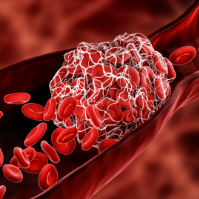
Chronic Limb Threatening Ischemia
Vascular Conditions
What is Chronic Limb-Threatening Ischemia? Ischemia (is-KE’me-ah) is a serious condition in which there is inadequate blood flow and oxygen to a specific part of the body. It can occur anywhere blood flows including the limbs, heart, brain, or intestines. It is generally caused by a narrowing or blockage of an artery. Ischemia is a severe condition that can cause tissue damage and loss of limbs. This condition will not improve on its own and requires appropriate medical attention. How does it occur? Ischemia can occur in any muscle group, organ, or tissue in the body. For example, in the lower extremities, ischemia can cause claudication in peripheral artery disease (PAD) or may cause chronic limb-threatening ischemia (CLTI) in severe cases. In the heart, ischemia is caused by coronary artery disease (CAD) and can cause angina (an-JI-nuh or AN-juh-nuh) or a heart attack; in the brain, ischemia may cause stroke or transient ischemic attacks (TIAs, or mini-strokes) and; in the intestines it is known as mesenteric ischemia and can cause severe abdominal pain. What are the symptoms of Ischemia? The symptoms of ischemia depend upon how quickly the blood flow is interrupted and where it occurs. In the limbs, early symptoms of ischemia may include claudication (pain, burning, or cramping in the muscles with exercise that goes away with rest). This can progress over time to CLTI, the reduction of blood flow to the affected extremity that results in severe pain or tissue loss. The pain is known as “rest pain” because it happens when resting or asleep. Usually, it wakes an individual up at night and can be relieved temporarily by hanging the leg over the bed or getting up to walk around. If the tissue of the limb has been affected, a non-healing sore or even gangrene may occur and the skin turns black; this requires immediate attention. Sudden onset of leg ischemia may cause severe pain, loss of pulses, coldness of the limb, paleness of the skin, and even leg weakness and loss of sensation. It also requires immediate medical attention. In the heart, ischemia may cause angina (chest pain with exertion that goes away with rest) or even a heart attack which requires immediate attention. In the brain, ischemia can cause a stroke and also requires immediate attention. In the intestines, if the blockage of the arteries supplying the intestines happens slowly, it results in chronic ischemia, which may cause abdominal pain after eating causing fear of food and unintended weight loss. In its acute sudden form, intestinal ischemia may cause bowel gangrene causing a sudden onset of severe abdominal pain that requires immediate medical attention. Risk factors of Ischemia? The risk factors are those for atherosclerosis (plaque buildup in the arteries): smoking, advanced age, high cholesterol, high blood pressure, diabetes, a family history of cardiovascular disease, sedentary lifestyle, and obesity. How is Ischemia diagnosed? Diagnosis of ischemia depends upon its location. Symptoms are the first clue, and your health care provider will ask you for detailed information on when and where the pain occurs, how often, and what relieves or exacerbates it. If the ischemia is in the lower extremities, you may be sent to a non-invasive vascular laboratory to have an ankle brachial index test (ABI), which tests the blood pressures in the legs. Depending upon those findings, you may be sent for a Duplex ultrasound imaging of the arteries or magnetic resonance arteriography (MRA) or computed tomography (CT). In some cases, particularly with chronic limb-threatening ischemia, you may be sent for an arteriogram, which is an x-ray of the arteries while dye is injected into the blood vessels. To diagnose a stroke, a picture of the brain with a head CT scan or a brain MRI is taken. Duplex ultrasound imaging, MRA, CT, or an arteriogram can also be used to assess or diagnose disease in the carotid arteries in the neck leading up to the brain. If heart ischemia is suspected you may receive a stress test. In severe cases you may be sent for coronary angiography. For mesenteric ischemia, imaging of the three vessels supplying the intestines is performed with ultrasound, CT, or MRA, followed by arteriography if necessary. How id Ischemia treated? Treatment of ischemia depends upon the location of the ischemia and its severity. However, for all forms of ischemia, controlling risk factors, especially smoking cessation, is essential! It may prevent progression of the ischemia and save your limb or your life. Treatment is focused upon getting more blood supply to the area of ischemia. This may be accomplished several ways: Medications Several medications may be prescribed to prevent further progression of the disease and to reduce the effect of contributing factors such as high blood pressure and cholesterol. Medications that fight infections, and pain medications may also be prescribed in certain cases. Exercise If one experiences claudication in the legs or angina in the heart, a supervised exercise routine may be prescribed. Regular exercise may also result in other benefits such as weight loss, lower blood pressure, lower cholesterol, and better control of diabetes. Surgery or Endovascular Procedures In many cases, an endovascular procedure may be performed within the artery using clot-busting drugs to dissolve clots (thrombolysis), a balloon to widen the artery (angioplasty), and/or a wire reinforced stent which remains inside the artery serving as a scaffolding to keep it open. In some cases, a bypass of the affected artery may be performed. Either a segment of leg vein or an artificial tube is attached surgically above and below the blockage to detour blood ow around the blocked area allowing the blood to reach the area of ischemia beyond the blockage. Examples of bypass procedures are leg artery bypass or coronary artery bypass grafting (also known as CABG or “open heart” surgery). Severe blockages in the carotid arteries that lead to ischemia may be treated with a surgical procedure known as carotid endarterectomy in which severe plaque is removed from the artery. Critical limb Ischemia is now chronic limb-threatening Ischemia Critical Limb Ischemia was renamed Chronic Limb-Threatening Ischemia (CLTI) in 2019. The name CLTI better reflects the broad range of patients with reduced blood flow that can delay wound healing and increase amputation risk. Source: https://vascularcures.org/chronic-limb-threatening-ischemia-clti/ Credit: Vascular Cures
Chronic Venous Insufficiency
SVS Vascular Conditions
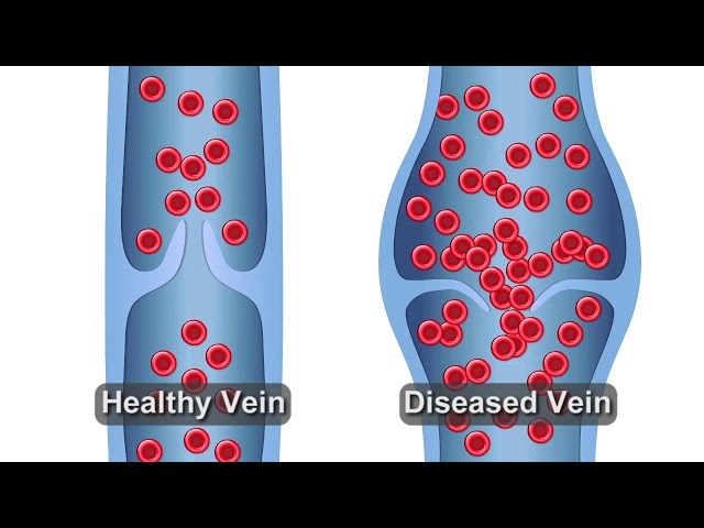
Chronic Venous Insufficiency Also known as Phlebitis, Post-Thrombotic Syndrome, Venous Insufficiency, Venous Leg Ulcer. If you have CVI, valves in your veins (usually in the leg or sometimes the arms) don't work, causing blood to pool in your legs and putting increased pressure on the walls of the veins. May be due to valve dysfunction (usually hereditary) or due to valve destruction after a deep vein thrombosis (DVT) or blood clot. Fairly common, usually chronic May affect up to 40% of the U.S population. More common in women (especially after multiple pregnancies) and in people who are middle-aged or older. Treatable - preventing complications is important Usually treated with exercise, compression stockings and weight loss if applicable. In some cases, may require vein ablation or vein stripping. Occasionally treated with angioplasty and stents. Preventing severe complications such as venous leg ulcers is key. Often recurs Even with very successful treatment, recurrence is common and you may need further care. Symptoms Most symptoms are mild, and not limb-threatening. Heaviness, swelling & pain You may feel heaviness in the affected limb, as well as swelling and pain. Sometimes there is a darkening of the skin. An open sore or ulcer may develop—if your leg is affected, usually on the inside of the ankle—and it may be difficult to heal. Varicose veins Enlarged and twisted veins close to the surface of the skin are a sign of a milder form of venous disease. You may feel burning, aching, heaviness and pain. Causes and Risks Vein valves become incompetent, especially when standing, for unknown reasons. A blood clot in a deep vein that causes your vein valves to fail and may completely block the vein. This causes greater swelling and can lead to tissue changes that may cause darkening of the skin, dermatitis or ulcers. Venous Insufficiency See: https://www.youtube.com/watch?v=CbHnb4TJqA8&t=4s Diagnosis - See a vascular surgeon You will be asked questions about symptoms and medical history, including questions about family members. The vascular surgeon will also perform a physical exam. Tests may be recommended Duplex ultrasound testing to look at your vein valves and see if they work. In cases where severe swelling occurs and is difficult to treat, a computer tomography (CT) scan of the venous system to look for vein narrowing or blockage in your abdomen. Treatments Most treatment is nonsurgical. The main goal is to prevent severe swelling and ulcers from developing. Compression garments relieve symptoms and aid ulcer healing. If superficial veins are affected, they may be treated through vein ablation or injection. If varicose veins develop and are close to the skin, they may be removed through superficial vein stripping, usually an outpatient procedure. If deep veins are affected, in severe cases angioplasty and stenting maybe recommended. In rare cases, surgical bypass may be required. Deep Vein Thrombosis Staying Healthy Maintain a normal weight Exercise regularly Wear compression garments Maintain good skin care If you have had a deep vein thrombosis (DVT), it is essential to stick with any anticoagulation medications you have been prescribed Source: https://vascular.org/your-vascular-health/vascular-conditions/chronic-venous-insufficiency
Chronic Venous Insufficiency (CVI)
Vascular Conditions
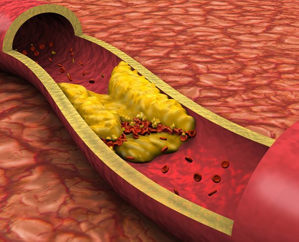
What is Chronic Venous Insufficiency (CVI)? Chronic venous insufficiency (CVI) is a common cause of leg pain and swelling, and is commonly associatedwith varicose veins. It occurs when the valves of the veins do not function properly, and the circulation of blood in the leg veins is impaired. CVI may affect up to 20% of adults. CVI can be caused by damaged valves in the veins or vein blockage. Both may be a result of deep vein thrombosis (DVT) or blood clots in the deep veins of the legs. If a clot forms in the superficial veins, there is a very low risk of DVT occurring. Over time, CVI may result in varicose veins, swelling and discoloration of the legs, itching and the development of ulcers near the ankles. Vein problems are among the most common chronic conditions in North America. In fact, more people lose work time from vein disorders than from artery disease. By the age of 50, nearly 40% of women and 20% of men have significant leg vein problems. Spider veins occur much more frequently in women. It is estimated that at least 20 to 25 million Americans have varicose veins. Causes The cause of CVI is related either to poorly functioning vein valves or blockage in the veins. Vein valves are designed to allow blood to flow against gravity from the legs back to the heart. When the valves fail to close properly, gravity wins and the flow reverses. This is called venous reflux. Vein valves may fail to close due to: Vein wall weakness that causes the vein to enlarge so that the valves cannot close A history of blood clots or deep vein thrombosis in the vein that damage the valve An absence of vein valves since birth Varicose veins are hereditary most of the time and generally occur in several members of the same family. Much less commonly, varicose veins develop after a trauma or injury. Regardless of the cause, defective valves cause a build up of pressure of the blood in the leg, leading to venous hypertension or high blood pressure in the vein. This may result in enlargement of the varicose veins and an increased likelihood of other symptoms such as swelling, skin changes and ulcers at the ankles or lower leg. Valve failure and venous reflux can also occur in the veins that are unseen, such as the saphenous veins (which run from the foot to the thigh) or in the deep veins. Reflux in these veins is often the underlying cause of painful varicose veins. Venous reflux is a condition that is progressive. If left untreated, it can worsen and cause more advanced symptoms of CVI. On occasion, the cause of the problem is not even in the legs, but is in the pelvis. Here, blockage of the veins may severely aggravate the symptoms of varicose veins, thus requiring separate treatment. Symptoms Varicose veins may be entirely symptom free and cause no immediate health problems. However, when varicose veins are associated with CVI, the most common sign is ankle and foot swelling that progress up the lower leg. Other skin changes in the lower leg that commonly occur include discoloration, eczema, scarring or hard, thickened skin and ulceration. When symptoms are present the most common are heaviness or fullness, aching, restlessness, tiredness, fatigue, pain, throbbing, burning, itching and muscle cramping. In advanced cases, breakdown of the skin may cause bleeding from varicose veins, and large varicosities may develop blood clots, a condition called superficial phlebitis or thrombophlebitis. If you experience any of these symptoms, talk with your doctor. Do not ignore these symptoms. Risk Factors The most important factors leading to the development of chronic venous insufficiency and varicose veins include: Family history Increasing age over 30 One or more blood clots in superficial or deep veins Female gender, although varicose veins occur nearly as commonly in men Multiple pregnancies Prolonged standing Heavy lifting Limited physical activity, high blood pressure and obesity have also been linked with the presence of varicose veins in women Diagnosis CVI is primarily diagnosed by physical examination. The accuracy of physical examination can be further improved with the aid of a hand-held instrument called a Doppler. This allows the examiner to listen to the blood flow. The most accurate and detailed test, however, is a venous duplex ultrasound exam. This provides an ultra-sound image or picture of the vein so that any blockage caused by blood clots or improper vein valve function can be detected. A CAT scan or MRI may be used to exclude other causes of leg swelling. These diagnostic tests are painless. Treatment The treatment of CVI involves both medical and surgical treatments: Diet and lifestyle Avoid prolonged standing or sitting Elevate the feet above the thighs when sitting and above the heart when lying down three to four times a day if possible to reduce swelling Structured exercise such as walking to strengthen calf muscles may improve calf muscle function Source: https://vascularcures.org/chronic-venous-insufficiency/ Credit: Vascular Cures
Compartment Syndrome VHC
SVS Vascular Conditions
Compartment Syndrome VHC Compartment syndrome can happen in any large muscular group within the body. However, it is most associated with the upper and lower extremities. Compartment syndrome occurs when swelling associated with muscle damage causes pressure within the tight sleeve of tissue that confines a muscle (the compartment). It results in insufficient blood supply to the tissue in that area. Symptoms Symptoms associated with compartment syndrome include Swelling in a limb after an injury or when blood flow has been interrupted and restored Pain in the calf or shin after a long period of running or walking Significant tenderness of leg muscles Causes and Risks There are two types of compartment syndrome, acute and chronic. Acute compartment syndrome is typically a surgical emergency arising from either an injury, such as a car crash, or when blood flow to a limb has been interrupted and then returned to normal, such as during surgery. The sudden increase in blood flow causes swelling that puts the nerves and muscles at risk of dying. Chronic compartment syndrome usually occurs in people who are exercising a great deal. After swimming, biking, running, or weightlifting, the muscles of the calf and shin become very painful to the point the person has to stop before they normally would. In addition, the patient will have significant leg muscle tenderness. Watch video: https://www.youtube.com/watch?v=oO9GqanB0s8 Diagnosis - Talk to a Doctor Active patients with unexplained calf and shin pain with exertion, especially if the muscles are tender to touch, should be evaluated for chronic compartment syndrome. Role of a Vascular Surgeon Anyone who has been told they have a limb threatening clot in an extremity should immediately seek a referral to a vascular surgeon. In one procedure, a vascular surgeon can both restore blood flow to the limb and release the compartment with a procedure called fasciotomy. Other doctors that may offer to remove clot but are not trained to perform the fasciotomy and thus a limb threatening delay in treatment may occur. Diagnosis Compartment syndrome can be a challenging diagnosis to make for both the acute and chronic forms. An accurate diagnosis requires a careful evaluation of the patient’s symptoms and examination of the affected extremity. For the acute form, time is of the essence and if there is uncertainty the vascular surgeon may place a needle into the compartment of the extremity to measure the compartment pressure. If it is elevated, then a fasciotomy may be necessary. However, most of the time the vascular surgeon can make the diagnosis without this step and will recommend a fasciotomy. For chronic compartment syndrome the vascular surgeon may order a CT scan, MRI scan or vascular doppler ultrasound as part of the evaluation. Treatments - Fasciotomy The specifics of the fasciotomy procedure can vary depending on the location and severity of the compartment syndrome: Incisions : The surgeon makes one or more incisions in the skin over the affected compartment. For acute compartment syndrome, long incisions may be required to adequately decompress the area. Fascia Opening : The fascia is then cut open to relieve the pressure. The muscle bulges through the opening, indicating that the pressure has been relieved. Monitoring and Closure : In cases of acute compartment syndrome, the wound might initially be left open (covered with a sterile dressing) to allow for continued swelling and to prevent re-accumulation of pressure. The skin can often be closed in a subsequent surgery, sometimes requiring skin grafts if the area cannot be closed directly due to swelling. In chronic compartment syndrome the incision is closed immediately. Nonsurgical Treatment For chronic compartment syndrome the vascular surgeon may recommend avoidance or discontinuation of the inciting activity if possible. Source: https://vascular.org/node/1847
Congenital Vascular Malformation
Vascular Conditions
What is Congenital Vascular Malformation? Birthmarks, Vascular Malformations and Anpmalies Vascular anomalies occur in barely 1% of all births. Yet, because of their rarity, their proper diagnosis and treatment is difficult, as most physicians do not see these problems often enough to become knowledgeable about their management. What are they? “Vascular anomalies” is an all-inclusive term for vascular malformations, vascular tumors and other congenital vascular defects. The more commonly used term, Congenital Vascular Malformation (CVM), implies abnormally formed blood vessels that one is born with. However, in spite of its redundancy, CVM is a popular term and it will be used here. Birthmarks occur on the surface of the body and are relatively easy to deal with. Other vascular malformations can develop from any type of blood vessel and develop in any part of the body, although most involve the extremities. They represent defects or development problems that occurred during embryonic growth. Depending on the state of development at the time this occurs, the result can involve arteries, veins, lymph vessels, or combinations of these. Birthmarks The difference between a CVM and a vascular tumor or hemangioma (the medical term), both of which are commonly called “birthmarks,” is very important to a child. Although they may initially appear the same, “all birthmarks are not the same.” Most birthmarks represent a superficial vascular malformation, consisting of abnormal collections of small blood vessels near the skin. Typically, this CVM type of “birthmark” does not go away nor does it enlarge, growing only at the same rate as the child. It thus maintains the same size and appearance indefinitely and is not a health threat, requiring no immediate treatment. Some birthmarks, due to their location, particularly around the face and neck or on some other exposed body part, may be considered unsightly. Fortunately, the characteristic reddish color coincides with the range of certain lasers that can be used for their removal. Another approach has been to cover them up by tattooing them a skin color. The other type of birthmark may appear the same at first, but is actually a vascular tumor or hemangioma. In contrast, this type grows rapidly in the months after its discovery, but then it “involutes” or gets progressively smaller. The majority disappear completely in a few years, leaving behind a patch of shrunken elastic skin. This regression normally is competed between two and eight years of age, but not all of them completely disappear. During their growth phase, these “juvenile hemangiomas” can be alarming, particularly if they grow in a critical location, such as those on the face impinging on the eye, nose, or mouth, in which case they may require treatment. However, most juvenile hemangiomas do not require treatment. Rather, the best advice is to do nothing but wait it out, giving it a chance to go away by itself. Both of these types of birthmarks, or the remnants of them, can be greatly improved in appearance by plastic surgery, but this is only occasionally needed and can usually be done after the child grows up. Arterial-Venous CVMS Other CVM are formed during early developmental stages when large connecting channels or shunts between future arteries and veins exist and for some unknown reason these artery-to-vein connections, or a cluster of them persist. Such connections are called arteriovenous fistulas (AVFs), or if there is a cluster of them they are called arteriovenous malformations (AVMs). These are potentially the most serious type of CVMs because in shunting blood from arteries to veins, they bypass the small vessels that make up the normal circulation beyond that point. This not only robs or steals blood that would otherwise pass through to more distant tissues and nourish them, but doesn’t allow for a gradual pressure drop from the high arterial pressure to the low pressure on the venous side. Thus, these AVFs represent a high flow short circuit, and depending on their size and location, they may force the heart to work harder. They may also cause poor circulation in the limb beyond the point of the AVF. In time, these AVFs tend to get bigger and have a greater effect on the circulation. For example, and AVF or AVM in an extremity can “steal” (reduce) blood flow to a foot or hand as much as a blocked artery would. Fortunately, AVFs located in the legs and arms are more common than elsewhere in the body, making them easier to deal with. Those involving pelvic vessels, or vessels to vital organs or the brain, can be extremely difficult to treat without injuring the organs or tissues surrounding them. Although AVFs make up only one third of all CVMs, they attract the most attention because of the serious problems they create, and are the CVMs most likely to need interventional treatment. Venous CVMS CVMs composed entirely of veins are the most common, comprising almost half of the total, and are of two basic types. The more primitive ones appear as thin-walled lakes in which venous blood collects. When they develop in groups or clusters, they may form a mass consisting of a collection of grape-like clusters of these venous lakes. This type usually does not affect the venous circulation which returns blood to the heart, but these malformations can be unsightly, cumbersome, or be the site of a type of blood clot (not the type that travel to the heart or lungs). They are not a serious threat, but may be worth treating if the mass is large and causes local problems, for example, interfering with walking. The other type of venous defect involves the large deep or central veins and often interferes with their function. Segments of major veins may be absent or narrowed. Some segments may be greatly widened and expanded (dilated), called a venous aneurysm. Treatment depends upon how severely they affect venous return or contribute to deep vein thrombosis (DVT). Most of the venous malformations involve only short venous segments and do not require treatment. Arterial CVMS CVMs of the arteries are the least common and responsible for only one 1-2% of the total. The most common arterial defect involves a segment that did not develop. The result is that a normal arterial segment is missing and instead blood flows through an undeveloped side channel or collateral artery, which persists rather than withers. Although this allows a bypass of the blockages, the enlarged bypassing segment often becomes more vulnerable to compression and injury, developing into an aneurysm or suddenly clotting off. The most common example of this is the so-called persistent sciatic artery. Symptoms of CVM When located in an extremity, CVMs may show up as a birthmark, a visible or palpable mass of blood vessels, may stimulate the development of collateral blood vessels in the form of varicose veins, or produce an enlargement of the limb or a lengthening of the limb by stimulating its bony growth centers. The localized masses may be of various sizes from small to huge. At their surface, the vessels may be vulnerable to injury and bleed, or may even break down and ulcerate. AVFs may cause “ischemic” pain, which is the medical term for pain that results when circulation is so restricted that the tissues and the nerves serving them do not get enough blood. How is CVM Diagnosed? Years ago, the only definitive way to evaluate blood vessel problems was by the injection of contrast dye that would make them visible on x-ray, called an angiogram. However, since most CVMs do not need treatment, or it is delayed until the need for treatment is more obvious, it is now rarely necessary to get angiograms as a first step. They may ultimately be needed, but only when an intervention is required, and even then are best obtained just before or at the time of treatment. Fortunately, great strides have been made in less invasive forms of vascular imaging. The localized superficial CVMs can often be initially studied by a form of ultra-sound imaging, called a color duplex scan. Larger mass lesions can best be studied by magnetic resonance imaging (MRI), which images in multiple planes (view angles), determines the anatomic extent of the malformation and importantly, whether its involvement of surrounding tissues (muscles, nerves, bones and joints) might preclude or complicate surgical treatment. CVM Treatment Options As a general rule, CVMs should be treated for specific indications: persistent pain, ulceration, bleeding, blood clots, obstruction of major vessels, causing progressive limb asymmetry by overgrowth, and for cosmetic indications or because the vascular mass is cumbersome and leads to a badly misshapen limb or interferes with extremity function in a mechanical way. Since most of the patients with the worst CVMs show up early in life, the timing of any intervention should be planned according to a child’s growth and development. Often it is better to delay operating on very young children, if possible. In the past, the only treatment for these vascular anomalies was surgical removal. However, of the CVMs that are significant and justify surgery, only 10-15% are removed. Removing even the simplest of these vascular malformations could lead to significant blood loss and is a surgical risk. Surgery may still be appropriate for localized, accessible lesions, but in the last few decades, techniques using catheters have been developed. Catheters are placed (usually through a groin vessel) into the lesions and the malformed vessels are blocked, or emblolized, with a variety of injectable particles, substances or devices such as polyvinyl foam, biological glues and absolute alcohol. These catheter embolization techniques can be used to control lesions without surgery. They can also shrink larger CVMs to make them more treatable by surgery. Laser therapy may also be effective for small, localized birthmarks (port wine stains). Patients with a rare venous malformation (Kleppel-Trenaunay Syndrome) of the limbs, frequently benefit from elastic garments and bandages used for compression of the large veins. After careful evaluation, surgery or less invasive therapy of the enlarged superficial veins can also be helpful. Source: https://vascularcures.org/congenital-vascular-malformation/ Credit: Vascular Cures