
Vascular Procedures
All Vascular Procedures
Angioplasty, Atherectomy and Stenting
Vascular Procedures
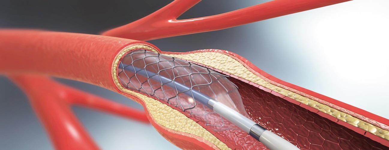
Angioplasty, Atherectomy and Stenting Arteries, blood vessels that carry oxygen-rich blood to all the parts of the body, are normally flexible and smooth on the inner side, but deposits of cholesterol, calcium, and fibrous tissue (plaque) can build up on the inner walls of the arteries, making them hard, stiff and narrow. This process of thickening and hardening of the arteries is called atherosclerosis. Atherosclerosis can reduce or completely stop the flow of blood through them, causing damage and pain to the part of the body that the arteries supply. Angioplasty is the surgical procedure that is performed to widen these blocked or narrowed arteries. During angioplasty, your surgeon inflates a small balloon within the narrowed artery to widen and improve the blood flow. Often, your surgeon may insert a stent, a tiny meshed tube to support the artery wall and keep the artery wide open Procedure Angioplasty is performed under local anaesthesia. A dye is injected into the body to view the blood flow through the arteries, and X-rays are taken. Using these X-ray images, your surgeon inserts a thin guide wire through a blood vessel in the groin area and extends it up to the blocked artery. A balloon catheter (thin tube) is then threaded through the guide wire to the narrowed artery. The balloon catheter has a deflated balloon at its tip, which is inflated at the site of blockage. The balloon widens the artery by compressing the plaque against the wall of the artery. Your surgeon may also insert a wire mesh tube called a stent along with the balloon catheter to help keep the artery open and prevent it from narrowing again. The balloon and the catheters are removed, while the stent is left in place to keep the artery wide open and allow free flow of blood. Post-procedure care Following the surgery, you will be able to go home the same day or may have to remain in the hospital for not more than 2 days. You will be able to start walking in 6 to 8 hours after the procedure. Risks & Complications Risks and complications following this procedure are rare. However, some specific complications for angioplasty are: bleeding from the catheter insertion site irregular heartbeat chest pain during procedure blood vessel damage from the catheter kidney damage from dye used with angiogram re-stenosis (re-accumulation of plaque or scar tissue causing narrowing or blockage of the coronary artery; occurs within 6 months) blood clots dislocation of stent Atherectomy Arteries, blood vessels that carry oxygen-rich blood to all the parts of the body, are normally flexible and smooth on the inner side. However, plaque (deposits of cholesterol, calcium, and fibrous tissue) can build up on the inner walls of the arteries. These plaques can make the arteries hard, stiff and narrow, reducing or completely blocking the flow of blood through them. Plaque can be removed by a procedure called atherectomy. Indications for Atherectomy Atherectomy is indicated for narrowed arteries that cannot be treated with: Angioplasty and stenting: procedure where a thin tube with a balloon and/or small mesh tube is inserted through a large artery in the leg to widen the blockage Bypass surgery: blood vessel re-directed to avoid arterial blockage Atherectomy is sometimes used as a complement to angioplasty and stenting where hardened plaques are first removed to an extent to allow the insertion of the balloon and stent, which then widen the artery. Procedure Atherectomy is performed under local anesthesia. A thin guide wire is threaded into the blocked artery with the help of a small needle that is inserted into a blood vessel in the groin area. This guides a catheter or a thin tube to the blockage, through which a dye is injected into the artery, and X-rays are taken to view the path of the artery. Your surgeon then inserts an atherectomy catheter attached with a blade, grinding bit, or laser filament at its tip through the guide wire to the narrowed part of the artery which is used to scrape away the plaque. Your surgeon may pass the catheter several times to cut away major parts of the hardened plaque. A collecting chamber or suction system at the tip of the catheter removes the plaque particles while it is being scraped off. Once the hardened plaque is removed, your surgeon inserts a stent, a tiny mesh tube, to support and keep the artery wall wide open, preventing it from narrowing again. Once the treatment is complete, the catheter is removed. Post-operative Care Following the procedure, you are advised to lie flat for some time. If bleeding occurs from the insertion site, you need to have pressure applied to stop the bleeding. You need to inform the nurse if you develop swelling, pain or bruising at the site of the catheter insertion. You will be asked to drink a lot of fluids to eliminate the dye injected into your body. Following the surgery, you will be able to go home the day after the atherectomy and you can return to your normal activities in a day or two, but you should refrain from engaging in certain strenuous activities. Risks & Complications The possible risks and complications of an atherectomy may include: Bleeding at the site of insertion Infections Re-blockage of the artery by plaque formation Heart attack due to closure of the artery, which requires emergency bypass surgery Damage or injury to the artery leading to loss of blood Clot formation in the leg or arm, which can lead to embolism (pieces of blood clot can break off and travel to the lungs or brain causing chest pain or stroke, respectively) Source: https://www.sydneyvascularsurgery.com.au/angioplasty-atherectomy-stenting.html
Arteriovenous Fistula Creation for Dialysis Access
Vascular Procedures
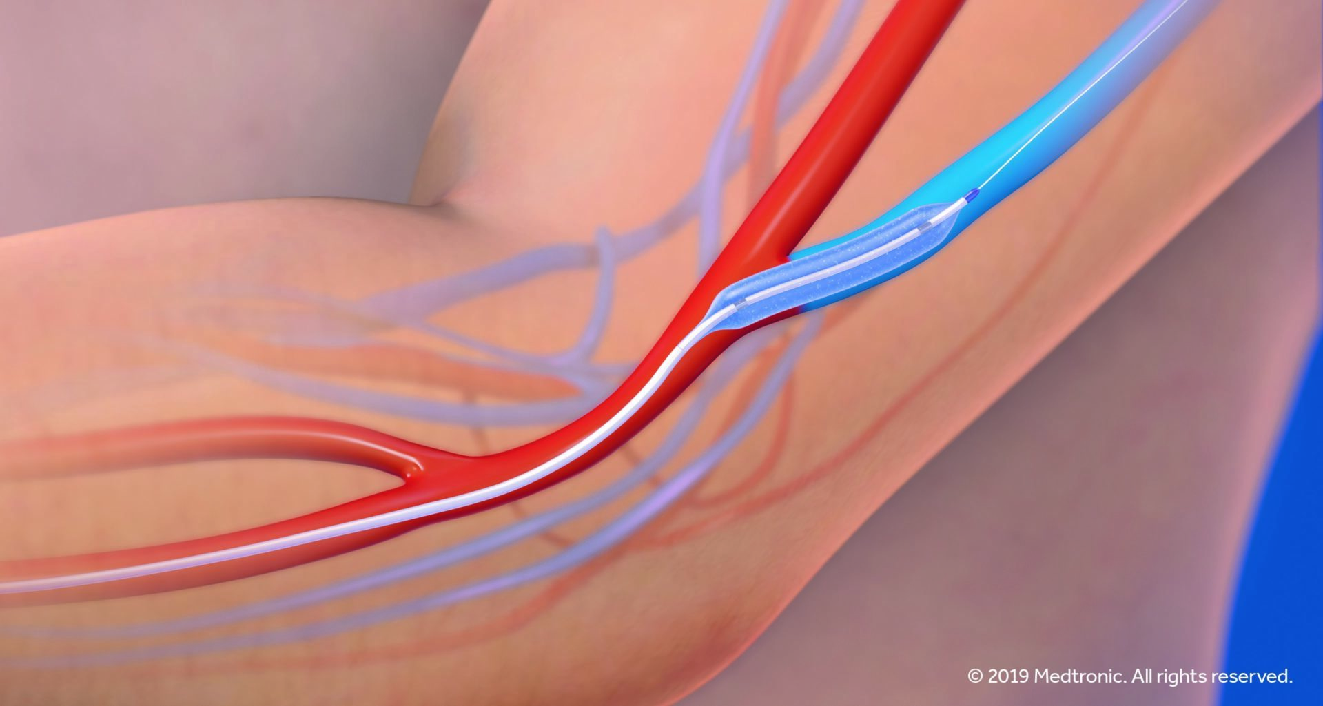
Arteriovenous Fistula Creation for Dialysis Access Haemodialysis is a procedure performed to manage patients suffering from kidney failure. A dialysis machine takes over the function of the diseased kidneys by filtering the blood of wastes. During dialysis, you will be attached to a machine called a dialyser through a thin tube. Your blood is then drawn into the machine for filtration through a needle inserted in your arm. The blood is circulated in the dialyser and the filtered blood is returned to the body through another tube. This is usually performed three times a week, so it requires easy access to the bloodstream each time. Vascular access is a surgically created large vein that provides easy access to the bloodstream and can withstand repeated needle insertions. There are two types of vascular accesses created for long-term use: Arteriovenous fistula: An abnormal connection surgically created between an artery and a vein. This allows the blood vessel to grow larger and have thicker walls, facilitating repeated puncturing. It provides good blood flow and is preferred for its lower rate of infection and clot formation, and greater longevity. Arteriovenous graft: A thin, flexible, synthetic tube (graft) is used to attach one end of an artery with another end of a vein. The graft is punctured to draw blood. The vascular access is created weeks or months before you can undergo haemodialysis. Source: https://www.sydneyvascularsurgery.com.au/arteriovenous-fistula-creation-for-dialysis-access.html

Carotid Endarterectomy
Vascular Procedures
Carotid Endarterectomy Carotid endarterectomy is a surgical procedure performed to remove plaque (deposits of fat) from the carotid arteries of the neck; the main blood vessels that supply blood to the brain. Indications Carotid endarterectomy is the most common treatment for carotid artery disease (CAD). The surgery is highly beneficial for people who have already had a stroke, or a transient ischemic attack (TIA). Surgery may also be recommended for individuals with severe narrowing of the carotid arteries without a history of any symptoms. Preparing for surgery Before having a carotid endarterectomy, you will receive specific instructions that help you prepare for the surgery. Most patients are on aspirin through the procedure but you will be asked to avoid certain medications before your surgery. A cerebral angiography, a diagnostic test that uses X-rays to produce detailed images of the blood vessels in your head, may be recommended before surgery. Procedure The aim of the surgery is to reduce the risk of stroke by shunting or rerouting the blood flow to the brain. A carotid endarterectomy often takes about 1 ½ to 2 hours and is usually performed by a vascular surgeon. The procedure is performed in a hospital under general or local anaesthesia with intravenous sedation. During the surgery, an incision is made over the neck to expose the narrowed carotid artery. A plastic tube is placed into the blood vessel, above and below the narrowing or blockage, to re-route the blood flow around the narrowed or blocked area. The artery is then opened and the plaque is removed. The plastic tube is then removed and the artery and skin incisions are closed. After surgery Recovery after a carotid endarterectomy is usually rapid and most patients are discharged home the next day after surgery. You may experience slight neck pain following the surgery, which is usually relieved with pain medications. Risks As with any type of surgery, there are some risks associated with a carotid endarterectomy which include infection, bleeding, blood clot formation, brain damage, and heart attack or stroke.Factors such as severity of the carotid artery disease, location of the blockage, previous history of stroke, and other health conditions may increase your risk of complications from this surgery. Carotid endarterectomy decreases the risk of brain damage and stroke. Adopting a healthy lifestyle can help prevent plaque build-up, blood clot formation, and other problems in the carotid arteries. Source: https://www.sydneyvascularsurgery.com.au/carotid-endarterectomy.html
Carotid Stenting
Vascular Procedures
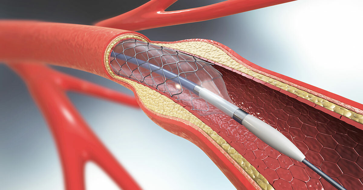
Carotid Stenting Carotid artery stenting is a minimally invasive procedure to open or clear the blocked carotid artery. It is performed along with carotid angiography, which uses X-ray images to check for the blocked carotid arteries. Carotid arteries are the arteries in the neck that supply blood to the brain. Carotid artery disease condition occurs by atherosclerosis, which is deposition of fatty material or plaque in the walls of carotid arteries. The fatty material thickens and becomes hard blocking the blood to flow within these arteries causing nerve cell death and stroke as a lack of oxygen to brain cells. You may be at higher risk of developing carotid artery disease if you have the following factors such as: Family history Inactive lifestyle or obesity Diabetes High blood pressure and high cholesterol Smoking Improper intake of medicines to control high blood pressure and high cholesterol Symptoms The symptoms do not develop at early stages of development. The most common symptoms include: Weakness and numbness in arm or legs on one side of body Slurred speech Headache Loss of vision in one of the eyes State of confusion Stroke Diagnosis If you notice any of the symptoms, then your cardiologist may perform physical examination and asks your medical history. Your doctor may request for diagnostic tests; the common tests include: Carotid duplex ultrasound – An ultrasound is placed over the carotid arteries to check for blood flow within the arteries Cerebral angiogram - A catheter is advanced into a large artery until the blocked artery in the neck area is reached. A contrast dye is then injected which helps to visualize the blocked artery by capturing images on an X-ray film. CT and magnetic resonance angiography (MRA) provides detailed images of the blood flow in the arteries. Procedure Carotid stenting is recommended by your cardiologist only if the conservative treatment such as medications and no improvement in changes to lifestyle habits. Carotid stenting is indicated in patients who are at increased risk of developing complications such as stroke while undergoing a surgical procedure called as carotid endarterectomy, to treat the blocked carotid arteries. During the stenting procedure, your surgeon performs angiogram procedure to identify the location of the blockage and how much the carotid arteries are blocked. During angiogram an incision is made in the groin or the thigh area and a catheter is pushed into the blood vessel until the blocked part is reached in the neck. A contrast dye is then injected into the artery and X-ray images are taken showing the location of the blockage. Now the surgeon will proceed for stenting procedure. While stenting procedure, a guide wire is inserted through the catheter and is advanced it to the blocked area. A clot protection device is placed. The device acts as a filter to trap the clots that escapes into the blood for circulation and helps to reduce the risk of stroke during the procedure. Another catheter is advanced into the blocked artery having a deflated balloon at one end through the guided wire into the blocked site. The balloon is then inflated which pushes the plaque against the artery walls and allows the blood to freely flow into this artery so as to reach the brain. The balloon can be deflated and inflated more than once to provide effective results. Your surgeon will then place a stent at the tip of the catheter before inserting it to the blocked site. When the balloon is inflated, the stent is pushed within the artery to help the artery to remain open. Some of the stents are coated with drugs to prevent formation of clots or prevent the artery to close. After the procedure, the balloon is deflated, the guide wire and catheter is withdrawn along with clot protection device. The stent remains in the artery and the incision is closed and wrapped with tight pressure bandage to prevent bleeding. Risks & Complications Complications related that may occur after carotid stenting procedure includes: Re-stenosis, re-accumulation of plaque narrowing or blocking the carotid artery again. Stroke - During the stenting procedure if the clot protecting device is not inserted, the blood clots may break and causes blockage in blood vessel. Stent may be repositioned. Source: https://www.sydneyvascularsurgery.com.au/carotid-stenting.html

Chronic / Non-healing Wound Management
Vascular Procedures
Chronic / Non-healing Wound Management Chronic non-healing wounds are the wounds that do not heal even after a few months or years, secondary to an underlying disease which may interfere with the normal healing process. Chronic wounds can be painful and can adversely affect the quality of life of the patient. Chronic wounds may lead to life threatening complications. Phases of wound healing The process of wound healing can be divided into four phases: Inflammatory phase: This involves a reflex vasoconstriction of the damaged blood vessels, in the wound, which reduces blood loss. Release of inflammatory mediators from the mast cells and damaged tissue causes inflammatory vasodilation which increases the permeability of the adjacent capillaries. Large molecules like fibrinogen escape into the wound area and form long sticky strands of fibrin, providing a framework for clot formation. It also acts as a physical barrier to compartmentalize the wound from the healthy tissue and prevents the spread of infection to the healthy tissue. Vasodilation also leads to localized increase in the flow of blood, increasing the supply of oxygen, nutrients and raw material for the repair of the injury. This occurs within the first 4 hours after the injury. Destructive phase: Within the next 48 hours’ white blood cells such as neutrophils and macrophages migrate to the wound area, attracted by the mediators released by the damaged tissue and facilitated by the increased permeability of the adjacent capillaries. They clear the dead tissue and any germs in the wound, a process similar to surgical debridement of wounds. Proliferation phase: Growth factors released from the macrophages and the damaged tissue attracts fibroblasts which synthesize and secrete collagen that aids in repair. This is followed by the formation of new blood vessels (angioneogenesis) and regeneration of epidermis (re-epithelialization). This phase starts about 2 to 3 days after the initial injury. Remodelling phase: In this phase the scar gradually decreases in size and finally fades away, acquiring the colour of the surrounding skin. This usually starts at about 3 weeks after the injury and may last up to a year or more depending on the size and nature of the wound. This phase involves myofibroblasts. Factors that delay wound healing Factors that delay wound healing may include conditions of the immediate wound environment (local factors) as well as conditions affecting the rest of the body (systemic factors). Blood supply to the wound is a crucial factor for wound healing. Thus, wound ischemia or conditions affecting the cardiovascular system can adversely affect wound healing. Immobility reduces the blood circulation and so reduces wound perfusion. Other factors that adversely affect wound healing include infection, presence of foreign body in the wound, poor hygiene, smoking, insufficient diet or malnutrition and psychological stress. Age also affects wound healing. Wound healing is slower in the elderly, as they have a reduced number of fibroblast in their tissues. This affects the formation of the three-dimensional scaffolding of collagen which is essential for healing. Re-epithelialisation and remodelling phase is also delayed. Moreover, presence of any underlying diseases such as diabetes mellitus, heart disease and peripheral ischemia may further hamper wound healing. Reduced mobility also increases the risk of pressure sore formation. Common Non-healing wounds Common non-healing wounds include: Pressure ulcers: Pressure ulcers are most commonly found over bony areas exposed to excessive pressure such as coccyx or tailbone, buttock, hip and heel. They occur due to reduced blood supply to the area, leading to the death of skin cells and formation of an ulcer. Pressure ulcers are more common in non-ambulatory patients. Diabetic foot ulcers: Diabetes causes nerve damage that can cause complete loss of sensation in the foot due to which minor foot injuries can lead to ulcers and infections and even gangrene. Diabetic foot is a term used for foot problems in patients with diabetes. Venous leg ulcers: These ulcers are commonly seen above the ankle. They occur due to insufficiency of the valves in the large leg veins which causes damage to the veins and the blood from the veins oozes out and collects under the skin. Treatment Treatment of chronic wounds involves debridement or wound cleaning and wound dressing. Sometimes special therapies such as hyperbaric oxygen therapy, vacuum assisted closure therapy and therapeutic ultrasound may also be used to improve wound healing. Large wounds may sometimes require skin grafts from another part of the body, usually the thigh, to cover them. Source: https://www.sydneyvascularsurgery.com.au/chronic-non-healin-ground-management.html
Endarterectomy
Vascular Procedures
Endarterectomy Normally, the arteries have a smooth surface inside to promote unobstructed flow of blood. With advancing age, a sticky plaque made up of cholesterol, calcium or fibrous tissue starts accumulating on the inner walls of your arteries. These plaques are called atherosclerotic plaques and the condition is termed as atherosclerosis. The deposition of plaque makes your arteries narrow and stiff. This can eventually lead to obstructed blood flow resulting in decreased oxygen supply to your organs and muscles. Endarterectomy is a surgical procedure to remove the atherosclerotic plaque from the narrowed artery. Endarterectomy minimizes the risk of complications that atherosclerosis may cause. Indication Endarterectomy is mainly performed if you have 50 to 70% narrowing of arteries. Endarterectomy has also proven to work better than medicines in people suffering from 70 to 99% blockage. This surgery is also recommended for the treatment of peripheral arterial disease, renal (kidney) artery disease, aortic arch conditions, aortoiliac occlusive disease, and visceral (intestines, spleen, and liver) artery disease. Pre-surgical care Your doctor may order an angiogram before the surgery. Angiogram is an invasive diagnostic test which gives a clear picture of the blood vessels. This helps your surgeon to ensure that it is safe to perform the surgery. You may be asked to discontinue some of your medicines before the surgery. Surgical procedure During the surgery, a small incision is made over the location of the artery to be treated and the artery is exposed. The blood flow is temporarily shunted (rerouted) with the help of a tube. The artery is cut opened and the plaque is carefully removed. A vein from the leg may be grafted onto the artery to repair the vessel. The shunt is then removed and the artery is stitched closed. The skin is finally sutured firmly. The complete surgery takes about 1 hour. Post-surgical care Soon after surgery, you will be moved to a recovery room for a short period of time. After this, you may be monitored in the intensive care unit for about 24 hours where you will be kept under close observation to watch development of any complications. You may be discharged after 1-3 days of hospital stay. You will be advised not to perform rigorous physical activities. There may be some pain in the area of surgery for the first 2 weeks. You can resume performing your normal daily activities within 1 or 2 weeks. Risks & Complications As seen with any surgical procedure, the complications that can occur due to endarterectomy are: Heart attack Stroke or transient ischemic attack Wound hematoma Intracerebral haemorrhage Cranial nerve deficits Repeated blockage of the artery Seizures (an uncommon complication) Infection Arrhythmias (irregular heart beat) Hypertension (high blood pressure) Airway obstruction from swelling Source: https://www.sydneyvascularsurgery.com.au/endarterectomy.html

Endovascular Stent Graft
Vascular Procedures
Endovascular Stent Graft Arteries are the blood vessels that carry oxygen-rich blood from the heart to all parts of the body. An aneurysm is a balloon like bulging or swelling in a weak area along the wall of an artery. An aneurysm can grow large, eventually bursting or rupturing causing internal bleeding and possibly death. An aortic aneurysm develops in the aorta (largest blood vessel in your body) and can be treated with an endovascular (inside a blood vessel) stent graft. Endovascular stent grafting is a new treatment method for abdominal aortic aneurysms and thoracic aortic aneurysms. It is a less invasive procedure when compared to an open surgery repair. Endovascular stent grafting involves use of an endovascular stent graft to reinforce the wall of the aorta and to prevent the damaged area from rupturing. Indication Your surgeon may recommend endovascular stent grafting if you are diagnosed with an abdominal aortic aneurysm or thoracic aortic aneurysm. Endovascular stent grafting is rarely used to treat aneurysms in other locations. Pre-surgical care Your surgeon may recommend for an MRI, CT scan or cerebral angiogram to evaluate the extent and location of the aneurysm and to decide your tolerability for surgery. Ultrasound and echocardiography can also help determine the size of the aneurysm. Angiography may be ordered to test the amount of damage or blockage of the blood vessels. In case of a ruptured aneurysm a cerebrospinal fluid sample may be tested as a pre-surgical procedure. You may also be advised to stop taking certain medication before surgery. Surgical procedure At the start of the procedure, you will be given regional anaesthesia or general anaesthesia depending on the need. A small incision is made in the leg near the groin. A catheter (thin tube) is inserted into an artery and advanced imaging technique is used to monitor delivery of the catheter carrying the stent graft to the area of the abdominal or thoracic aortic aneurysm. The surgeon fastens the stent graft in correct position and removes the delivery catheter. The stent graft placed inside the aorta prevents the aneurysm from bursting, allowing blood to flow freely without pushing on the weakened area of the artery. Post-surgical care Normally, you will have to stay in hospital for 2 to 3 days after surgery. After the first day of recovery you will be permitted to eat and walk. You will be asked to avoid driving and lifting heavy weights for almost 4 to 6 weeks or until the physician approves. You will be advised to return for a follow-up visit after the first few weeks. An imaging test may be recommended after a year to monitor the blood vessels. Risks & complications The complications of endovascular stent grafting include: Infection Leaking of blood around the graft Graft fracturing Movement of the graft away from the desired location Blockage of the blood flow through the graft Source: https://www.sydneyvascularsurgery.com.au/endovascular-stent-graft.html
Renal Transplant and Vascular Access
Vascular Procedures
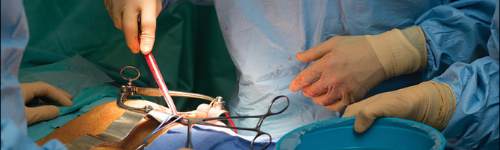
Renal Transplant & Vascular Access What is Renal Failure & Dialysis Access? Renal failure is a serious condition when the kidneys - organs that filter toxins from the body - are unable to remove waste products from the blood stream. The toxins and waste products then accumulate in the blood stream, causing uraemia. The process of renal failure is slow going, so initially, the kidneys may continue to do some filtering work and are able to compensate for a mild degree of failure. Eventually though, the kidneys are unable to keep the pace and will degrade into chronic renal failure. Dialysis is the method in which the blood is filtered. It is like having an artificial kidney. There are two methods of dialysis - peritoneal and haemodialysis. Haemodialysis is a filtering of the blood using a dialysis machine. For this, access to the blood via the veins and arteries is required. Dialysis Access is the term for methods used to access the veins and arteries to get a complete flow of blood through the dialysis machine. A highly successful way to access the veins and arteries for dialysis is through the creation of an arteriovenous fistula. What causes Renal Failure? Some of the causes of renal failure include: Diabetes - even if it is well managed, diabetes can cause kidney damage. High blood pressure Glomerulonephritis - swelling or inflammation of the tiny filtering units (nephrons) in the kidney Polycystic kidney disease - an inherited condition Urinary reflux - urine flows backwards into the kidneys, causing sclerosis. Medications - some drugs such as lithium and cyclosporin can cause kidney failure. There are factors that may increase your risk of chronic kidney disease that may lead to renal failure, such as: Having diabetes Having high blood pressure Being obese Over 60 years of age Having a family history of end-stage kidney disease or hereditary kidney disease in a first or second degree relative Having established heart problems (heart failure or a past heart attack) or have had a stroke Smoking Having a history of acute kidney injury Being of Maori or Pacific origin Being of Aboriginal or Torres Strait Islander origin What are the signs/symptoms of Renal Failure? The early stages of kidney disease are usually asymptomatic. Worryingly, advanced stages of kidney disease can still be asymptomatic, even when up to 90% of kidney function is gone. This is why it is important to detect any renal degeneration early, to prevent kidney failure. Once symptomatic, kidney disease symptoms can include: Tiredness Loss of appetite Difficulty sleeping Headaches Lack of concentration Shortness of breath Nausea and vomiting Changes in the amount and number of times urine is passed Changes in the appearance of urine or blood in the urine Puffiness in the legs and ankles Pain in the kidney area These symptoms can be caused by other conditions, but if you are in a high-risk group for kidney disease, you should speak with your doctor. How will Renal failure, and Dialysis, affect my health? Renal failure is a serious condition. Patients with chronic renal failure need lifelong dialysis or a kidney transplant to stay alive. There is an important distinction between kidney disease and kidney failure. If you have kidney disease, it does not mean that you will develop kidney failure. As mentioned earlier, your kidneys can continue to work at less than 100% for a long time, and if you have kidney disease, you will likely not experience any symptoms until the kidneys are very damaged. What are the stages of Renal Failure and Dialysis Access? Renal failure can be acute (happening very quickly and a medical emergency) or chronic (happening over many months, or years). Chronic kidney disease is when you have lost over 30% of your kidney function. It is a gradual process that happens over the course of many years. Chronic kidney failure is end stage chronic kidney disease. The kidney no longer functions and dialysis, or a kidney transplant, is required. Because this process takes a long time, you will likely visit a vascular surgeon and other specialists, who together would have developed a treatment plan in consultation with you. I think I have Renal Failure – What should I do? Prevention is key to avoiding renal failure. It is much easier to monitor kidney function throughout your life, than to suddenly find yourself in need of a new kidney! It is almost impossible to know if you have kidney disease or failure without monitoring, until it is very advanced. If you find a reduction in kidney function during the course of monitoring tests, it is ideal to visit a GP and seek a referral to a Vascular Surgeon. The SVS Vascular Surgeons can review your medical history, assess your lifestyle and present treatment plans that work best, including information about the most appropriate dialysis access method. What if I don’t have my Renal Failure treated? This is unfortunately very simple to answer. Without treatment, either with dialysis or a kidney transplant, you will not survive. What treatment options does SVS offer for Renal Failure and Dialysis Access? Sydney Vascular Surgery offers two treatment options for renal failure: Renal Transplant Arteriovenous Fistula creation & surveillance Both treatments are surgical, life-extending and do require monitoring for the remainder of your life. Renal transplant A renal transplant is a kidney transplant, either from a living or deceased (cadaveric) donor. It is considered a treatment, not a cure, providing a potentially longer and more active life free of dialysis. At Sydney Vascular Surgery, Dr Vikram Puttaswamy has spent many years working closely with the Royal North Shore Hospital Renal Department and Renal Transplant team and has experience in performing living, cadaveric and Paired Kidney Exchange (PKE) transplants . You can have a transplant if you are medically suitable. If the transplant is from a living donor, the operation can be done before you need dialysis. The average wait for a kidney from a deceased donor is about four years. Preparing for organ donation surgery is a complex and time consuming process. It is important and it is life-changing. By the time you see a vascular surgeon in the presence of chronic kidney disease or kidney failure, you are already accustomed to seeing many specialists and you will already have an excellent idea of what type of transplant you are to be treated with. If you are visiting this site today and you are not a registered organ donor, we strongly encourage you to reconsider. If you would like more information please visit Donate Life . Arteriovenous fistula creation and management An arteriovenous fistula is a direct connection between an artery and a vein that is created in a surgical operation. The connection is made so that the vein enlarges over a period of weeks, with the aim to make the vein larger, and the vein wall tougher, to be able to withstand future dialysis needle punctures. The vein increases in size because arterial blood at a higher pressure is now flowing through the vein. The vein wall toughens (matures) over a few weeks or months. When dialysis is required needles are inserted into the vein and connected to a dialysis machine. The blood leaves the body through one needle, is filtered through the dialysis machine, and returns to the body through the other needle. An arteriovenous fistula can be created using native veins (called ‘autogenous fistulae’) or with the use of a synthetic material such as gortex or PTFE (called a ‘PTFE forearm loop’). Autogenous fistulae are referred as they are likely to work for longer and require less maintenance. Arteriovenous fistulas are most commonly created in the arm or leg. Arm fistulas are much more common than in the leg. It is preferable to locate the fistulae as low on the arm as possible, closest to the hand, and on the non-dominant arm. Source: https://www.sydneyvascularsurgery.com.au/renal-transplant.html
Surgical Aneurysm Repair
Vascular Procedures
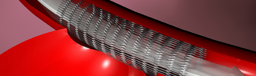
Surgical Aneurysm Repair An aneurysm is a balloon like enlarged and weakened area (caused due to blood pressure) on the wall of an artery. An aneurysm can cause serious complications when it increases in size and ruptures or when blood clots block the blood flow. Aneurysms can be safely treated by surgical repair. In surgical aneurysm repair, your surgeon repairs or removes an aneurysm to promote normal blood flow. Indication Aneurysms occur mostly in the aorta, the largest artery in your body running from your heart through the chest and abdomen. Surgical aneurysm repair is indicated for the treatment of abdominal aortic aneurysm, an aneurysm that occurs in your abdomen and for thoracic aortic aneurysm, an aneurysm that occurs in your chest. This method is also employed for the treatment of aneurysm in arteries located in your thigh, spleen, knee, kidneys, liver, and stomach. Pre-surgical care At first, your physician will obtain your medical history and perform a physical examination. You may be ordered to get some tests done such as a CT scan, duplex ultrasound and angiography. Your doctor might ask you not to eat or drink anything for 8 hours before the surgery. Your doctor will also discuss with you whether you need to reduce the dose or stop taking certain medications. Surgical procedure You will be given anaesthesia to block the pain during the surgery. Your vascular surgeon will make a small incision in your skin and muscle over the artery with the aneurysm. The surgeon exposes the aneurysm site and clamps the artery above the aneurysm to stop the blood flowing through the area. The aneurysm is cut opened to remove the blood clots and plaque deposits. Your surgeon may insert a graft that is attached just above and below the aneurysm to bypass the weakened area. Alternately, your surgeon may attach a fabric patch to the wall of the artery to strengthen it and decrease its size. Or, a tiny metal or plastic coil is inserted using a catheter into the area of the aneurysm. These methods all help to facilitate normal blood flow. Post-surgical care You will be shifted to the intensive care unit after surgery and you will be monitored continuously till you recover. You may need to stay in the hospital for 7-10 days until your physician considers your health to have improved satisfactorily. You will be advised not to lift anything and to follow the diet instructions strictly. Risks & complications The complications of surgical aneurysm repair include: Breathing problems Heart problems Colon problems Kidney problems You can improve your lifestyle after surgical aneurysm repair by quitting smoking, having healthy eating habits, exercising regularly and maintaining a normal body weight. Source: https://www.sydneyvascularsurgery.com.au/surgical-aneurysm-repair.html
Surgical Bypass
Vascular Procedures
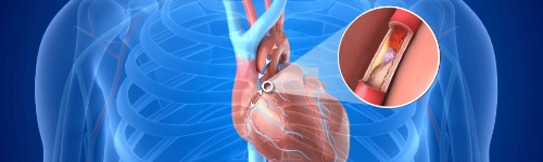
Surgical Bypass Normally, the arteries have a smooth surface inside to promote unobstructed flow of blood. A sticky plaque made up of cholesterol, calcium or fibrous tissue starts building up on the inner walls of your artery, as you age. The accumulation of these atherosclerotic plaques in the artery makes it stiff and hard. Eventually the blood flow is blocked due to atherosclerosis resulting in decreased supply of oxygen to your organs and muscles. This condition can be successfully treated with surgical bypass. Surgical bypass involves treating your narrowed arteries by rerouting around a section of the artery that is blocked. Surgical bypass is usually performed in patients who are not good candidates for angioplasty or who have found angioplasty to be ineffective. It is a good treatment option for atherosclerosis. Before surgery, your physician checks your general health, medical history, and symptoms. You will have to undergo a physical examination and you may be ordered for a blood test to determine your cholesterol levels. In addition to this, duplex ultrasound, magnetic resonance angiography or computerized tomographic angiography may be recommended to determine the extent of blockage. During bypass surgery, you will receive anaesthesia so that you do not feel any pain. Your surgeon will make an incision over the artery that is blocked. The clamps will be placed at each end of the blocked section of artery. The graft is then sutured in place. The doctor will make sure that the alignment is correct without any leakage, blood flows properly and the graft is working fine. Your surgeon then stitches the incision firmly. After surgery, you might have to stay for 3-10 days in the hospital for monitoring. You should contact your physician immediately if you develop a fever, a cold painful arm or leg, or if the incision area becomes red or swollen. You may have to follow a strict diet and take prescribed medications regularly. You can resume your physical activities only as per your doctor’s advice. You will be asked to meet your doctor for follow-up visits. As with any surgical procedure, surgical bypass carries some minor risks such as swelling, bleeding at the incision site, wound healing problems and infection. Source: https://www.sydneyvascularsurgery.com.au/surgical-bypass.html
Thoracic Outlet Surgery
Vascular Procedures
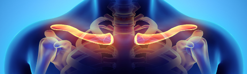
Thoracic Outlet Surgery The thoracic outlet is a small passageway leading from the base of the neck to the armpit and arm. This small area contains many blood vessels, nerves and muscle. When this passageway becomes compressed the condition is termed as thoracic outlet syndrome. This rare condition is characterized by burning pain in the neck and shoulder, numbness and tingling of the fingers, and a weak hand grip. Thoracic outlet syndrome generally occurs within the age group of 20 to 60 years and is more common in females than in males. There are 3 types of thoracic outlet syndrome namely neurogenic thoracic outlet syndrome, arterial thoracic outlet syndrome and venous thoracic outlet syndrome. Thoracic outlet syndrome can result due to injury, tumours that press nerves, poor posture that compresses nerves, weight lifting, anatomical defects such as an elongated C7 transverse process, anomalous tissue overgrowth, upper thoracic neurovascular compression, costocoracoid tendon, subclavian muscle hypertrophy and so on. The symptoms of thoracic outlet syndrome may include numbness and pain in the neck, shoulder and arm, tingling or burning sensation, weakness, limited range of movement of arms, and swelling or redness of your arm, or colour changes to the arm or hand. A proper diagnosis is very essential for the condition of thoracic outlet syndrome. To evaluate your condition, your doctor will perform a physical examination and collect your medical history. You may be advised to have a chest X-ray, MRI or CT scan. Your doctor might also recommend for blood tests and electromyogram evaluation. Nerve conduction velocity study may be suggested to check the conduction of electrical signals in the nerves. The treatment of thoracic outlet syndrome involves both non-surgical and surgical approach. Non-surgical Treatment Physical therapy- Exercises can decrease the pressure on the nerves and blood vessels and strengthen the muscles surrounding the shoulder. Maintaining proper posture can help you stand, sit and walk straight. Non-steroidal anti-inflammatory medications- can reduce the pain and swelling. Weight loss- reducing your weight, if you are considered over weight. Lifestyle changes- avoid strenuous activities and other activities that increase the risk or symptoms. Surgical Treatment If nonsurgical treatment does not alleviate your symptoms, your doctor may advise surgery. Surgery may involve removing a portion of an abnormal first rib or releasing a muscle that joins the neck and chest. The surgery will depend on the cause of the compression. Source: https://www.sydneyvascularsurgery.com.au/thoracic-outlet-surgery.html
Thrombolytic Therapy
Vascular Procedures
Thrombolytic Therapy The normal process of blood clotting or coagulation occurs when platelets clump with other blood components to form a gel. Generally, clotting after an injury avoids excessive bleeding, but clots formed in the blood vessels of the body may block the blood flow in vital organs such as the lungs, heart or brain creating a life threatening situation which requires emergency management. Thrombolytic therapy breaks up fatal blood clots in the blood vessels by administering either clot-dissolving medications or inserting a catheter through the blood vessels to mechanically break the clot. Indications Thrombolytic therapy is most commonly indicated to treat a blood clot in the blood vessels of your brain such as in ischemic stroke. Other indications for therapy include: Pulmonary embolism (clot in the lung artery) Deep vein thrombosis (clot in the deep veins of legs) Clot in the heart (heart attack) Clot in the arteries of arms or legs Blocked dialysis access catheter or bypass graft Pre-procedural Preparation Before thrombolytic therapy is undertaken, your doctor will order tests to confirm that you are a candidate to receive the therapy. You may be asked to stop medications that increase the risk of bleeding and other complications. You will have to undergo angiography with a contrast agent so that the internal structure of your blood vessels and location of the clot can be viewed before the therapy. Procedure Thrombolytic therapy can be delivered by inserting a catheter in the vein and guiding it towards the clot. Your doctor will first clean and shave the site of catheter insertion on the skin and numb the skin with local anaesthetic agent. A small incision or puncture is made in the skin and the catheter is inserted along with the contrast agent to track the blood vessels. X-rays will be taken to locate the clot in the blood vessels. Thrombolytic medications will be injected through the catheter near the location of clot to break or dissolve the clot. Your doctor will monitor the breaking of the clot with help from X-ray images. The procedure can vary from several hours to even days before the clot is fully dissolved. Once the clots have dissolved, your doctor will check for satisfactory range of the blood’s ability to coagulate and remove the catheter. The site of puncture will be compressed for 20 minutes to stop bleeding. Sometimes, your doctor may perform a mechanical procedure to treat the clots. Small devices such as a suction cup, fluid jet, rotating device or ultrasound device is placed on the end of the catheter tip to physically break the clot. Post-procedural care Following the procedure, you will have to rest for several hours to prevent bleeding from the punctured site. Inform your doctor if you experience fever, pain, or swelling and discolouration of the limb. You may be prescribed medicines, diet or physical therapy after an emergency thrombolytic therapy. Risks & Complications As with any procedure, thrombolytic therapy involves certain risks and complications. They include: Bleeding at the puncture site Low blood pressure Rarely, bleeding in the brain Source: https://www.sydneyvascularsurgery.com.au/thrombolytic-therapy.html