
Heart Procedures
All Heart Procedures
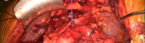
Aortic Root Surgery
Heart Procedures
Aortic Root Surgery Overview Aortic root surgery is a procedure to treat an enlarged section of the aorta, also known as an aortic aneurysm . The aorta is the large blood vessel that carries blood from the heart to the body. The aortic root is where the aorta and the heart connect. Aortic aneurysms near the aortic root may be due to an inherited condition called Marfan syndrome. Other causes include heart problems present at birth, such as a bicuspid aortic valve. Why it's done Aortic root surgery is done to prevent: A rupture of the aorta. A tear between the layers of the aorta's wall, called an aortic dissection. Stretching of the aortic valve. The risk of these events depends on the size of the aortic root and cause of enlargement. Aortic root surgery is recommended for those at high risk. How you prepare Before aortic root surgery, talk to your health care provider about the best treatment or surgery for your condition. Also discuss the benefits and risks of the surgery. What you can expect Ascending aortic root aneurysm procedure There are several types of aortic root surgery, including: Aortic valve and root replacement. This procedure also is called composite aortic root replacement. A surgeon removes part of the aorta and the aortic valve. Then, the surgeon replaces the section of the aorta with an artificial tube, called a graft. The aortic valve is replaced with a mechanical or biological valve. Anyone who has a mechanical valve needs to take blood-thinning medicine, also called blood thinners, for life to prevent blood clots. Blood thinners also are called anticoagulants. Valve-sparing aortic root repair. A surgeon replaces the enlarged section of the aorta with a graft. The aortic valve stays in place. In one technique, the surgeon sews the valve inside the graft. If you have another heart condition, your surgeon may be able to treat it at the same time as aortic root surgery. Source: https://www.mayoclinic.org/tests-procedures/aortic-root-surgery/about/pac-20383396

Atrial Fibrillation Ablation
Heart Procedures
Overview Atrial fibrillation ablation is a treatment for an irregular and chaotic heartbeat called atrial fibrillation (A-fib). It uses heat or cold energy to create tiny scars in the heart to block the faulty electrical signals and restore a typical heartbeat. Atrial fibrillation ablation may be used if medications or other treatments don't work. Sometimes it's the first treatment for certain patients. Atrial fibrillation ablation is most often done using thin, flexible tubes called catheters inserted through the veins or arteries to the heart. Less commonly, ablation is performed during cardiac surgery. Types AV node ablation Maze procedure Why it's done Atrial fibrillationOpen pop-up dialog box Atrial fibrillation ablation is done to reset the heart's rhythm. A health care provider may recommend this type of ablation if you have A-fib symptoms, including a fast, fluttering heartbeat, that hasn't improved with medication or other treatments. Risks Possible atrial fibrillation ablation risks include: Bleeding or infection at the site where the catheters were inserted Blood vessel damage Heart valve damage New or worsening irregular heartbeats (arrhythmias) Slow heart rate that could require a pacemaker to correct Blood clots in the legs or lungs (venous thromboembolism) Stroke or heart attack Narrowing of the veins that carry blood between the lungs and heart (pulmonary vein stenosis) Damage to the kidneys from contrast dye used during the procedure Death, rarely Discuss the risks and benefits of atrial fibrillation ablation with your health care provider to decide whether it's right for you. How you prepare Your health care provider may order several tests to get more information about your heart condition before your atrial fibrillation ablation. You'll need to stop eating and drinking the night before your procedure. Your care provider will tell you how or if you should continue any medications before atrial fibrillation ablation. What you can expect Before Atrial fibrillation ablation is done in the hospital. A care provider will insert an IV into your forearm or hand and give you a medication called a sedative to help you relax. The amount of sedation needed for the procedure depends on your specific arrhythmia and other health conditions. You may be fully awake or lightly sedated, or you may be given general anesthesia (fully asleep). During Catheter insertion points for cardiac ablationOpen pop-up dialog box One of the following ablation techniques is used to create small scars in the heart and block the irregular heartbeats: Heat (radiofrequency energy) Extreme cold (cryoablation) You may feel some minor discomfort when the catheter is moved into your heart or when the dye is injected and when energy is being delivered. If you have severe pain or shortness of breath, let your care providers know. The procedure usually takes three to six hours. Complicated procedures may take longer. After Following your procedure, you'll be moved to a recovery area to rest quietly to prevent bleeding from the site where the catheters were inserted into the blood vessels. Care providers will continuously monitor your heartbeat and blood pressure to check for complications. Depending on your condition, you may be able to go home the same day as your procedure. If you go home the same day, plan to have someone drive you. You may feel a little sore after your procedure, but the soreness shouldn't last more than a week. You'll usually be able to return to your daily activities within a few days after atrial fibrillation ablation. Results Most people see improvements in their quality of life after cardiac ablation. But there's a chance that the irregular heartbeats may return. If this happens, the procedure may be repeated or your health care provider might recommend other treatments. The procedure hasn't been shown to reduce the risk of a stroke. Blood thinners may be needed to reduce stroke risk. Source: https://www.mayoclinic.org/tests-procedures/atrial-fibrillation-ablation/about/pac-20384969
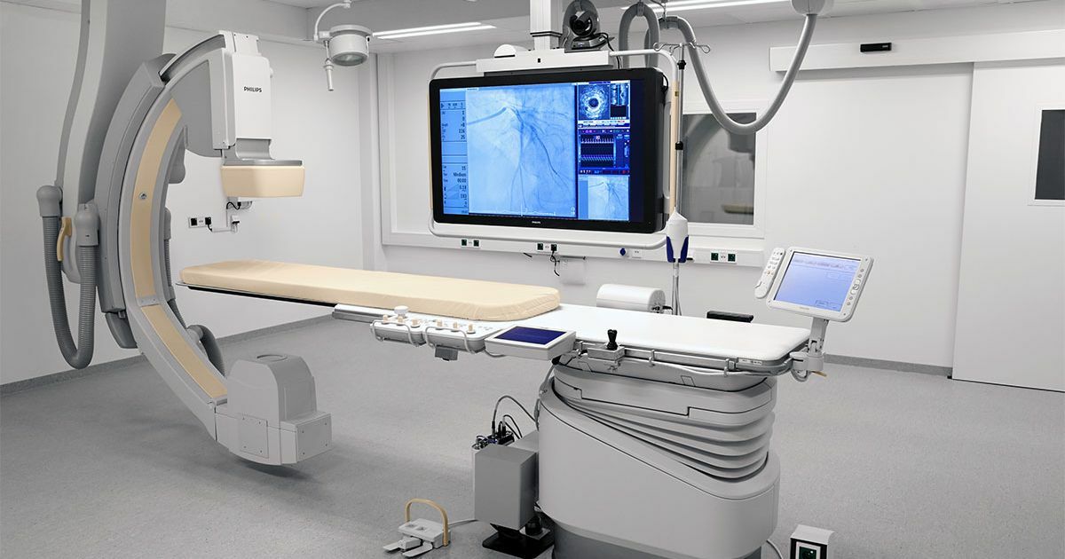
Cardiac Catheterization
Heart Procedures
Cardiac Catheterization Interventional cardiologists are highly skilled at diagnosing and treating heart problems with cardiac catheterization. Whether it is a diagnosis for a heart disorder or an emergency treatment for a guest experiencing chest pain or a heart attack, they can help with your treatment options. Specialists in catheterization diagnose treat many heart problems. Unlike surgery, which requires large incisions, cardiac catheterization involves inserting a catheter, a thin flexible tube, through a small nick in the groin or wrist and guiding it to the arteries of the heart. This usually means you have less discomfort, heal faster and get back to your life more quickly than with open heart surgery. Safety and quality are carefully monitored by a panel of physicians who review every angioplasty procedure done in hospital. All of these procedures are also reported to a national quality registry. Cardiac catheterization procedures Interventional cardiologists perform a variety of heart procedures in the cath lab to treat or manage your heart condition. Cardiac catheterization procedures include: Angioplasty and stenting to treat coronary artery disease Angioplasty and stenting for peripheral artery disease (PAD) affecting the legs or kidneys Balloon dilation and stent placement to treat coarctation of the aorta Balloon valvuloplasty and transcatheter aortic valve replacement (TAVR) to treat aortic valve stenosis and balloon valvuloplasty to treat mitral valve stenosis Carotid angioplasty and stenting Chronic total occlusion (CTO) coronary intervention Closure of periprosthetic valve leaks Clot removal with aspiration device (thrombectomy) Treatment to repair a leaking mitral valve Nonsurgical closure of ostium secundum atrial septal defect (ASD) or patent foramen ovale Percutaneous mitral valve replacement and repair, a minimally invasive procedure to treat mitral valve disease Plaque removal with a rotating device (atherectomy), laser device, and cutting balloon Pulmonary artery angioplasty to open lung arteries blocked with chronic clots and help improve pulmonary hypertension Pulmonary vein intervention to treat veins blocked after an ablation procedure Robotic coronary angioplasty and stenting Septal ablation to treat hypertrophic cardiomyopathy Transcatheter insertion of coil or occluder to treat patent ductus arteriosus (PDA) Transcatheter mitral valve repair (TMVR) to treat mitral regurgitation Transcatheter pulmonary valve replacement (TPVR) for congenital heart problems with the pulmonary valve Percutaneous ventricular assist devices Heart conditions treated include Procedures conducted in the cath lab to diagnose your condition may include: Coronary, carotid, renal, peripheral and other forms of angiography Coronary anatomy and physiology assessment with intravascular ultrasound (IVUS), optical coherence tomography (OCT), or fractional flow reserve (FFR) Endomyocardial biopsy Evaluation of complex valvular disease Left and right heart hemodynamic assessment Constriction and restriction studies Source: https://www.bswhealth.com/the-heart-hospital/treatments-and-procedures/cardiac-catheterization
Cardiothoracic Surgery
Heart Procedures
Experts in open-heart surgery, robotic cardiac surgery and minimally invasive heart surgery Cardiothoracic surgeons are doctors who operate on the heart, lungs, esophagus, diaphragm, blood vessels and other organs in the chest. Cardiothoracic surgeons combine experience, surgical skill and advanced facilities with a team-based, compassionate approach to deliver patient-centered care. Cardiothoracic surgeons design a treatment approach for you, from diagnosis to cardiothoracic surgery to after-care, including cardiac rehabilitation when necessary. Highly skilled specialists have extensive experience in heart and chest surgery, with surgeons that are board-certified in thoracic surgery, general surgery and surgical critical care. Search for providers that have a 3-star rating from The Society of Thoracic Surgeons (STS) for its patient care and outcomes in four categories: aortic valve replacement surgery (AVR), AVR plus CABG surgery, Mitral Valve Repair and Replacement (MVRR) and Coronary Artery Bypass Grating (CABG). The 3-star rating, which is one of the most sophisticated and highly regarded overall measures of quality in healthcare. Hospitals like Baylor Scott & White The Heart Hospital – Plano are among the most elite for cardiac surgery in the United States and Canada. Heart conditions treated with Cardiothoracic Surgery include Cardiothoracic surgeons are doctors who operate on the heart, lungs, esophagus, diaphragm, blood vessels and other organs in the chest. Aneurysms (abdominal aortic and thoracic aortic aneurysms) Congenital heart disorders Coronary artery disease Esophageal cancer Gastroesophageal reflux and other disorders Heart failure Heart rhythm disorders Heart valve disorders Lung cancer Tumors of the chest wall, chest and mediastinum Cardiothoracic Surgery treatments include Adult congenital heart surgery Coronary artery bypass graft (CABG) surgery, including: Traditional surgery Robotic minimally invasive direct coronary artery bypass (MIDCAB) Thoracoscopic CABG surgery Major aortic surgery and endovascular aortic surgery, including aortic dissection, traumatic aortic rupture and thoracic aortic aneurysm treatment Radiofrequency ablation of lung cancers Thoracic surgery for lung cancers, esophageal cancer and chest injury repair Transcatheter aortic valve replacement (TAVR) Heart valve surgery, including: Minimally invasive aortic valve replacement Port access mitral valve repair Robotic approaches Ventricular assist device implantation, including: Destination therapy Ventricular assist device (VAD) Source: https://www.bswhealth.com/the-heart-hospital/treatments-and-procedures/cardiothoracic-surgery
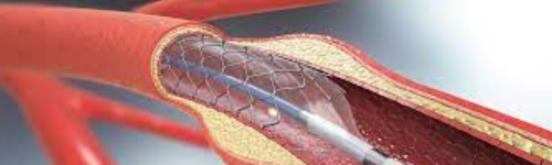
Coronary Angioplasty and Stents
Heart Procedures
Coronary Angioplasty and Stents Overview Coronary angioplasty (AN-jee-o-plas-tee), also called percutaneous coronary intervention, is a procedure used to open clogged heart arteries. Angioplasty uses a tiny balloon catheter that is inserted in a blocked blood vessel to help widen it and improve blood flow to the heart. Angioplasty is often combined with the placement of a small wire mesh tube called a stent. The stent helps prop the artery open, decreasing its chance of narrowing again. Most stents are coated with medication to help keep the artery open (drug-eluting stents). Rarely, bare-metal stents are used. Angioplasty can improve symptoms of blocked arteries, such as chest pain and shortness of breath. Angioplasty is also often used during a heart attack to quickly open a blocked artery and reduce the amount of damage to the heart. Why it's done Development of atherosclerosis Angioplasty is used to treat the buildup of fatty plaques in the heart's blood vessels. This buildup is a type of heart disease known as atherosclerosis. Angioplasty may be a treatment option for you if: You have tried medications or lifestyle changes but these have not improved your heart health. You have chest pain (angina) that is worsening. You have a heart attack. Angioplasty can quickly open a blocked artery, reducing damage to your heart. Angioplasty isn't for everyone. Depending on the extent of your heart disease and your overall health, your doctor may determine that coronary artery bypass surgery is a better option than angioplasty for you. You may need coronary artery bypass surgery if: The main artery that brings blood to the left side of your heart is narrow Your heart muscle is weak You have diabetes and multiple severe blockages in your arteries In coronary artery bypass surgery, the blocked part of your artery is bypassed using a healthy blood vessel from another part of your body. Risks Although angioplasty is a less invasive way to open clogged arteries than bypass surgery is, the procedure still carries some risks. The most common angioplasty risks include: Re-narrowing of your artery. When angioplasty is combined with drug-eluting stent placement, there's a small risk the treated artery will become clogged again. The risk of re-narrowing of the artery is higher when bare-metal stents are used. Blood clots. Blood clots can form within stents even after the procedure. These clots can close the artery, causing a heart attack. It's important to take aspirin in combination with clopidogrel (Plavix), prasugrel (Effient) or another medication that helps reduce the risk of blood clots exactly as prescribed to decrease the chance of clots forming in your stent. Talk to your doctor about how long you'll need to take these medications. Never stop taking these medications without discussing it with your doctor. Bleeding. You may have bleeding in your leg or arm where a catheter was inserted. Usually this simply results in a bruise, but sometimes serious bleeding occurs and may require a blood transfusion or surgical procedures. Other rare risks of angioplasty include: Heart attack. Though rare, you may have a heart attack during the procedure. Coronary artery damage. The coronary artery may be torn or ruptured during the procedure. These complications may require emergency bypass surgery. Kidney problems. The dye used during angioplasty and stent placement can cause kidney damage, especially in people who already have kidney problems. If you're at increased risk, your doctor may take steps to try to protect your kidneys, such as limiting the amount of contrast dye and making sure that you're well hydrated during the procedure. Stroke. During angioplasty, a stroke can occur if plaques break loose when the catheters are being threaded through the aorta. Blood clots also can form in catheters and travel to the brain if they break loose. A stroke is an extremely rare complication of coronary angioplasty. Blood thinners are used during the procedure to reduce the risk. Abnormal heart rhythms. During the procedure, the heart may beat too quickly or too slowly. These heart rhythm problems are usually short-lived, but sometimes medications or a temporary pacemaker is needed. How you prepare Before a scheduled angioplasty, your doctor will review your medical history and do a physical exam. You may need to have some tests, including a chest X-ray, electrocardiogram and blood tests, before your procedure. Your doctor will also perform an imaging test called a coronary angiogram to see if the arteries to your heart are blocked and if they can be treated with angioplasty. If your doctor finds a blockage during your coronary angiogram, he or she may decide to perform angioplasty and stenting immediately after the angiogram while your heart is still catheterized. Your doctor will give you instructions to help you prepare. Your doctor may instruct you to adjust or stop taking certain medications before angioplasty, such as aspirin, nonsteroidal anti-inflammatory drugs (NSAIDs) or blood thinners. Be sure to tell your doctor about all medications you take, including herbal supplements. Usually, you'll need to stop eating or drinking six to eight hours before an angiography. Take approved medications with only small sips of water on the morning of your procedure. Gather all of your medications to take to the hospital with you, including nitroglycerin, if you take it. Arrange for transportation home. Angioplasty usually requires an overnight hospital stay, and you won't be able to drive yourself home the next day. What you can expect During the procedure Angioplasty is performed by a heart specialist (cardiologist) and a team of specialized cardiovascular nurses and technicians in a special operating room called a cardiac catheterization laboratory. Angioplasty is performed through an artery in your groin, arm or wrist area. General anesthesia isn't needed. You'll receive a sedative to help you relax, but you may be awake during the procedure depending on how deeply you are sedated. You'll receive fluids, medications to relax you and blood-thinning medications (anticoagulants) through an IV catheter in your hand or arm. Your heart rate, pulse, blood pressure and oxygen level will be monitored during the procedure. Your doctor will prepare the area in your leg, arm or wrist with an antiseptic solution and will place a sterile sheet over your body. Your doctor will use a local anesthetic to numb the area where a very small incision will be made. A small, thin guidewire is then inserted into the blood vessel. With the help of live X-rays, your doctor will thread a thin tube (catheter) through your artery. Contrast dye is injected through the catheter once it is in place. This allows your doctor to see the inside of your blood vessels and identify the blockage on X-ray images called angiograms. A small balloon with or without a stent at the tip of the catheter is inflated at the site of the blockage, widening the blocked artery. After the artery is stretched, the balloon is deflated and the catheter is removed. If you have several blockages, the procedure may be repeated at each blockage. Angioplasty can take up to several hours, depending on the difficulty and number of blockages and whether any complications arise. You might feel pressure in the area where the catheter is inserted. You may also feel some mild discomfort when the balloon is inflated and your artery is stretched, but typically you shouldn't feel any sharp pain during the procedure. Coronary artery stent Stent placement Most people who have angioplasty also have a stent placed in their blocked artery during the same procedure. A stent, which looks like a tiny coil of wire mesh, supports the walls of your artery and helps prevent it from re-narrowing after angioplasty. Here's what happens during a stent placement: The stent, which is collapsed around a balloon at the tip of the catheter, is guided through the artery to the blockage. At the blockage, the balloon is inflated and the spring-like stent expands and locks into place inside the artery. The stent stays in the artery permanently to hold it open and improve blood flow to your heart. In some cases, more than one stent may be needed to open a blockage. Once the stent is in place, the balloon catheter is deflated and removed. More X-ray images (angiograms) are taken to see how well blood flows through your newly widened artery. Most stents implanted during an angioplasty are drug coated. The medication in the stent is slowly released to help prevent future plaque buildup and the re-narrowing of the blood vessel. After your stent placement, your doctor will prescribe medications, such as aspirin, clopidogrel (Plavix), ticagrelor (Brilinta) or prasugrel (Effient), to reduce the chance of blood clots forming on the stent. After the procedure If you had a nonemergency procedure, you'll probably remain at the hospital overnight while your heart is monitored and your medications are adjusted. You generally should be able to return to work or your normal routine the week after angioplasty. When you return home, drink plenty of fluids to help flush your body of the contrast dye. Avoid strenuous exercise and lifting heavy objects for at least a day afterward. Ask your doctor or nurse about other restrictions in activity. Call your doctor's office or hospital staff immediately if: The site where your catheter was inserted starts bleeding or swelling You develop pain or discomfort at the site where your catheter was inserted You have signs of infection, such as redness, swelling, drainage or fever There's a change in temperature or color of the leg or arm that was used for the procedure You feel faint or weak You develop chest pain or shortness of breath Blood thinners It's important that you closely follow your doctor's recommendations about your treatment with blood-thinning medications — aspirin and clopidogrel (Plavix), prasugrel (Effient) or similar medications. Most people who have undergone angioplasty with or without stent placement will need to take aspirin indefinitely. Those who have had stent placement will need a blood-thinning medication, such as clopidogrel, for six months to a year. If you have any questions or if you need any other type of surgery, talk to your cardiologist before stopping any of these medications. Results Coronary angioplasty greatly increases blood flow through the previously narrowed or blocked coronary artery. Chest pain generally should decrease. You may be better able to exercise. Having angioplasty and stenting doesn't mean your heart disease goes away. You'll need to continue healthy lifestyle habits and take medications as prescribed by your doctor. If you have symptoms similar to those you had before coronary angioplasty, such as chest pain or shortness of breath, contact your doctor. If you have chest pain at rest or pain that doesn't respond to nitroglycerin, call 911 or emergency medical help. To keep your heart healthy after angioplasty, you should: Quit smoking Lower your cholesterol levels Eat a healthy diet that is low in saturated fat Maintain a healthy weight Control other conditions, such as diabetes and high blood pressure Get regular exercise Take medications as prescribed by your doctor Successful angioplasty also means you might not have to undergo coronary artery bypass surgery, a more invasive procedure that requires a longer recovery time. Source: https://www.mayoclinic.org/tests-procedures/coronary-angioplasty/about/pac-20384761

Coronary Artery Bypass Surgery
Heart Procedures
Coronary Artery Bypass Surgery Overview Coronary artery bypass surgery Coronary artery bypass surgery creates a new path for blood to flow around a blocked or partially blocked artery in the heart. The surgery involves taking a healthy blood vessel from the chest or leg area. The vessel is connected below the blocked heart artery. The new pathway improves blood flow to the heart muscle. Other names for this surgery are: Coronary artery bypass grafting. CABG — pronounced "cabbage." Coronary artery bypass graft surgery. Heart bypass surgery. Coronary artery bypass surgery doesn't cure the heart disease that caused a blockage, such as atherosclerosis or coronary artery disease. But it can reduce symptoms such as chest pain and shortness of breath. The surgery, commonly called CABG, may reduce the risk of heart disease-related death. Why it's done Coronary artery bypass surgery is done to restore blood flow around a blocked heart artery. The surgery may be done as an emergency treatment for a heart attack, if other immediate treatments aren't working. Your health care provider might recommend coronary artery bypass surgery if you have: A blockage in the left main heart artery. This artery supplies a lot of blood to the heart muscle. Severe narrowing of the main heart artery. Severe chest pain caused by narrowing of several heart arteries. The narrowing reduces blood flow to the heart even during light exercise or at rest. More than one diseased heart artery and your lower left heart chamber doesn't work well. A blocked heart artery that can't be treated with coronary angioplasty. This less-invasive treatment uses a balloon on the tip of a thin tube, called a catheter, to widen the artery. A small coil called a stent is typically used to keep the artery open. An angioplasty with or without a stent that hasn't worked. For example, an artery narrowed again after stenting. Source: https://www.mayoclinic.org/tests-procedures/coronary-bypass-surgery/about/pac-20384589

Electrocardiogram (ECG or EKG)
Heart Procedures
Electrocardiogram (ECG or EKG) Overview Electrocardiogram An electrocardiogram records the electrical signals in the heart. It's a common and painless test used to quickly detect heart problems and monitor the heart's health. An electrocardiogram — also called ECG or EKG — is often done in a health care provider's office, a clinic or a hospital room. ECG machines are standard equipment in operating rooms and ambulances. Some personal devices, such as smartwatches, offer ECG monitoring. Ask your health care provider if this is an option for you. Types Holter monitor Implantable loop recorder: A heart monitoring device Why it's done An electrocardiogram is a painless, noninvasive way to help diagnose many common heart problems. A health care provider might use an electrocardiogram to determine or detect: Irregular heart rhythms (arrhythmias) If blocked or narrowed arteries in the heart (coronary artery disease) are causing chest pain or a heart attack Whether you have had a previous heart attack How well certain heart disease treatments, such as a pacemaker, are working You may need an ECG if you have any of the following signs and symptoms: Chest pain Dizziness, lightheadedness or confusion Heart palpitations Rapid pulse Shortness of breath Weakness, fatigue or a decline in ability to exercise The American Heart Association doesn't recommend using electrocardiograms to assess adults at low risk who don't have symptoms. But if you have a family history of heart disease, your health care provider might suggest an electrocardiogram as a screening test, even if you have no symptoms. If symptoms tend to come and go, they may not be detected during a standard ECG recording. A health care provider might recommend remote or continuous ECG monitoring. There are several different types. Holter monitor. A Holter monitor is a small, wearable device that records a continuous ECG, usually for 24 to 48 hours. Event monitor. This portable device is similar to a Holter monitor, but it records only at certain times for a few minutes at a time. You can wear it longer than a Holter monitor, typically 30 days. You generally push a button when you feel symptoms. Some devices automatically record when an irregular rhythm is detected. ECG on medical helicopter A person receives an ECG while on a helicopter for medical transport. Source: https://www.mayoclinic.org/tests-procedures/ekg/about/pac-20384983
Electrophysiology
Heart Procedures
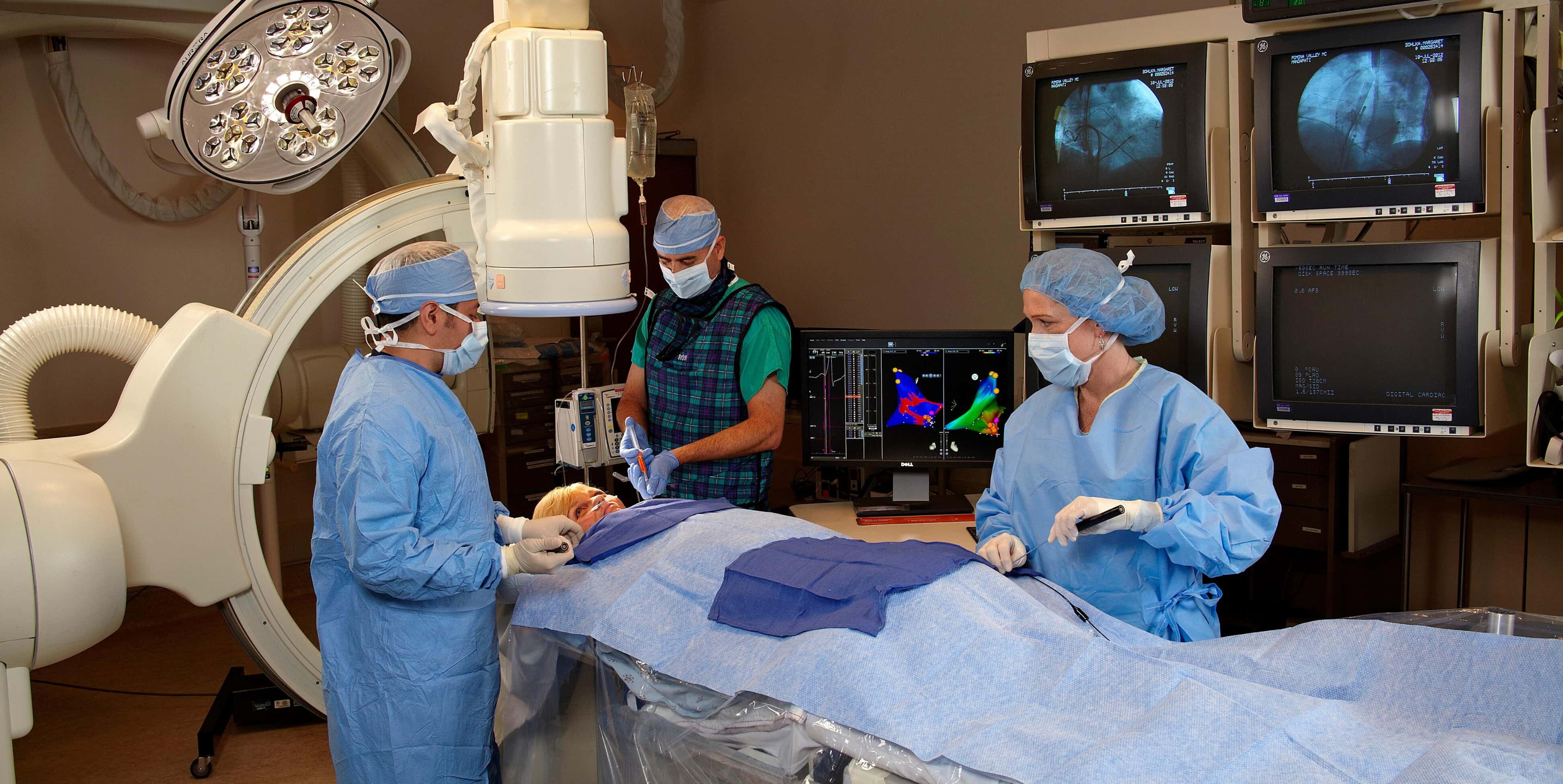
Electrophysiology Electrophysiology heart conditions include Arrhythmias, also known as heart rhythm disorders, occur when there is a change in the normal rate or rhythm of the heartbeat. They are generally caused by abnormalities in the heart's electrical system. Cardiac arrhythmias can be complex medical conditions with effects ranging from annoying to life-threatening. Heart arrhythmias include: Atrial fibrillation Atrial flutter Atrioventricular block (also known as heart block) Long QT syndrome Pacemaker/ICD malfunction Paroxysmal supraventricular tachycardia (PSVT) Rhythm disorders related to heart failure Premature atrial contraction Sick sinus syndrome Sinus tachycardia Sinus bradycardia Sudden cardiac death prevention Ventricular fibrillation Ventricular tachycardia Diagnostic and treatment services Although some arrhythmias can be managed by medication, others may require surgery. Electrophysiologists are experts at both traditional open surgeries and minimally invasive procedures. Minimally invasive surgeries use small incisions and precisely directed catheter-based tools to treat heart rhythm disorders. Heart patients usually benefit from these less-invasive procedures through shorter recovery time, smaller incisions and better outcomes. Atrial fibrillation management Cardiac resynchronization therapy Diagnostic heart monitoring Holter monitor Event monitoring Remote mobile cardiac outpatient telemetry monitoring Electrophysiology study Lead management and extraction Maze procedure Pacemaker and bi-ventricular pacemaker implantation Radiofrequency ablation Sudden cardiac death assessment and evaluation Supraventricular tachycardia (SVT) ablation Syncope evaluation and management, including tilt table testing Ventricular fibrillation and tachycardia management and ablation Zero-fluoroscopy ablation Source: https://www.bswhealth.com/the-heart-hospital/treatments-and-procedures/electrophysiology
Emergency Cardiac Care
Heart Procedures
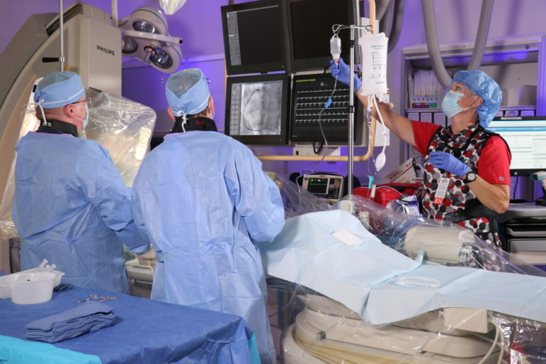
Emergency Cardiac Care Hospital based, emergency cardiac care departments are 24-hour, seven-days-a-week, accredited chest pain centers with board-certified emergency room physicians and cardiologists on the medical staff, as well as specially trained nurses. Heart physicians are highly skilled in treating heart attacks and other cardiovascular emergencies. *Door-to-balloon time is an initiative that measures how much time it takes to get a patient who is experiencing chest pain or a heart attack from the door of the ER to the operating room to open an affected artery. American Heart Association CPR and ECC The AHA is the leader in resuscitation science, education, and training, and publisher of the official Guidelines for CPR and ECC . Millions of healthcare providers and others trust the AHA for their lifesaving training, and 100% of the AHA's profits go back into supporting its lifesaving mission. Hands-Only CPR Our hands can do so many things, the most important of which may be saving someone’s life. The power is in YOUR hands. See: https://cpr.heart.org/en/cpr-courses-and-kits/hands-only-cpr An Emergency Lifesaving Procedure CPR – or Cardiopulmonary Resuscitation – is an emergency lifesaving procedure performed when the heart stops beating. Immediate CPR can double or triple chances of survival after cardiac arrest. What is CPR? CPR: Cardiopulmonary Resuscitation Science-based Guidelines The AHA develops science-based CPR guidelines and is the leader in first aid, CPR, and AED training. All recommendations below are based on the AHA Guidelines Update for CPR and Emergency Cardiovascular Care (ECC) . CPR – or Cardiopulmonary Resuscitation – is an emergency lifesaving procedure performed when the heart stops beating. Immediate CPR can double or triple chances of survival after cardiac arrest. The American Heart Association invites you to share our vision: a world where no one dies from cardiac arrest. Every year, 350,000 people die from cardiac arrest in the United States. Big number. Bigger opportunity. With your help, we can bring that number down to zero. Immediate CPR can double or triple chances of survival after cardiac arrest. Why Is CPR Important? Keeping the blood flow active – even partially – extends the opportunity for a successful resuscitation once trained medical staff arrive on site. Chain of Survival CPR is a critical step in the AHA’s Chain of Survival. The term Chain of Survival provides a useful metaphor for the elements of the ECC systems concept. The 6 links in the adult out-of-hospital Chain of Survival are: Recognition of cardiac arrest and activation of the emergency response system (calling 9-1-1 in the US) Early CPR with an emphasis on chest compressions Rapid defibrillation Advanced resuscitation by Emergency Medical Services and other healthcare providers Post-cardiac arrest care Recovery (including additional treatment, observation, rehabilitation, and psychological support) A strong Chain of Survival can improve chances of survival and recovery for victims of cardiac arrest. Automated External Defibrillators (AED) AEDs can greatly increase a cardiac arrest victim's chances of survival. The AHA offers a two-page guide on how to implement an AED program at a company or organization. To minimize the time to defibrillation for cardiac arrest victims, deployment of AEDs should not be limited to only trained people (although training is still recommended). Learn more about how the use of AEDs can dramatically boost survival of cardiac arrest patients. Implementing an AED Program (PDF) How is CPR Performed? There are two commonly known versions of CPR: For healthcare providers and those trained: conventional CPR using chest compressions and mouth-to-mouth breathing at a ratio of 30:2 compressions-to-breaths. In adult victims of cardiac arrest, it is reasonable for rescuers to perform chest compressions at a rate of 100 to 120/min and to a depth of at least 2 inches (5 cm) for an average adult, while avoiding excessive chest compression depths (greater than 2.4 inches [6 cm]). For the general public or bystanders who witness an adult suddenly collapse: compression-only CPR, or Hands-Only CPR. Hands-Only CPR is CPR without mouth-to-mouth breaths. It is recommended for use by people who see a teen or adult suddenly collapse in an out-of-hospital setting (such as at home, at work, or in a park). Hands-Only CPR consists of two easy steps: Call 9-1-1 (or send someone to do that) Push hard and fast in the center of the chest Learn About Hands-Only CPR For a visual comparison of Hands-Only CPR vs. Conventional CPR, view the CPR Guide Infographic (PDF). About High-Quality CPR High-quality CPR should be performed by anyone - including bystanders. There are five critical components: Minimize interruptions in chest compressions Provide compressions of adequate rate and depth Avoid leaning on the victim between compressions Ensure proper hand placement Avoid excessive ventilation Even Children Can Perform Successful CPR A recent study tested sixth graders and their capacity to use Hands-Only CPR to save lives. The study found that the majority of children could perform CPR in the correct location and at the appropriate compression rate, making this a viable group to train to help save lives. In fact, the AHA is dedicated to training the next generation of lifesavers through its CPR in Schools program. We led the charge to make CPR training a high school graduation requirement in 34 states – and counting! Learn more and get resources and training kits designed specifically for schools. The Gender Difference: Men vs. Women According to a study released by the Resuscitation Science Symposium , men are more likely to receive bystander CPR in public locations compared to women. Women and men receive similar CPR assistance within the home, but in public 45 percent of men received assistance compared to only 39 percent of women. Men were 1.23 times more likely to receive bystander CPR in public, and their chance of survival was 23 percent higher compared to women. So why the discrepancy? It could come down to anatomy and a bystander being comfortable enough to perform CPR on a woman. Heart Attack vs. Cardiac Arrest The Difference You Should Know For more information on Cardiac Arrest vs. Heart Attack, view the infographic. What is Cardiac Arrest? Occurs when the heart malfunctions and stops beating unexpectedly. Cardiac arrest is an “ELECTRICAL” problem. What is a Heart Attack? Occurs when blood flow to the heart is blocked. A heart attack is a “CIRCULATION” problem. Find A Course Find a Course Near You If you are interested in taking a course to learn the lifesaving skills of CPR, first aid, and AED, use the Find A Course Tool to locate a Training Center near you. Use the Find A Course Tool Try a Blended Learning Course Many AHA lifesaving training courses are available online via ShopCPR . Courses that involve only cognitive learning can be completed entirely online. For courses that teach CPR, students must complete an in-person skills practice and testing session with an AHA Instructor after they complete the online portion. The Instructor will evaluate the student's skills and upon successful completion of both portions of the course, the student will receive a course completion card, valid for two years. See more information on blended learning and eLearning training . CPR-Related Inquiries: 1-877-AHA-4CPR or 1-877-242-4277 International Inquiries Global Web Support (email) Source: https://cpr.heart.org/en/resources/what-is-cpr Source: https://www.bswhealth.com/the-heart-hospital/locations/plano/emergency-cardiac-care
Heart Scan (coronary calcium scan)
Heart Procedures
Heart Scan (coronary calcium scan) A heart scan, also known as a coronary calcium scan, is a specialized X-ray test that provides pictures of your heart that can help your doctor detect and measure calcium-containing plaque in your arteries. Plaque inside the arteries of your heart can grow and restrict blood flow to the muscles of your heart. Measuring calcified plaque with a heart scan may allow your doctor to identify possible coronary artery disease before you have signs and symptoms. Your doctor will use your test results to determine what you need — medication or lifestyle changes — to reduce your risk of a heart attack or other heart problems. Why it's done Your doctor may order a heart scan to get a better understanding of your risk of heart disease or if your treatment plan is uncertain. A heart scan uses a specialized X-ray technology called multidetector row or multislice computerized tomography (CT). The scan creates multiple images that can show any plaque deposits in the blood vessels. A heart scan provides an early look at levels of plaque. Plaque is made up of fats, cholesterol, calcium and other substances in the blood. It develops gradually over time, long before there are any signs or symptoms of disease. These deposits can restrict the flow of oxygen-rich blood to the muscles of the heart. Plaque may also burst, triggering a blood clot that can cause a heart attack. When is a heart scan used? A heart scan may help guide treatment if you have a low to moderate risk of heart disease or if your heart disease risk isn't clear. Your doctor can tell you if you might benefit from having a heart scan based on your risk factors. A heart scan may also help motivate people at moderate risk to make important lifestyle changes and follow treatment plans. Source: https://www.mayoclinic.org/tests-procedures/heart-scan/about/pac-20384686
Heart Transplant
Heart Procedures
Heart Transplant A heart transplant is an operation in which a failing heart is replaced with a healthier donor heart. Heart transplant is a treatment that's usually reserved for people whose condition hasn't improved enough with medications or other surgeries. While a heart transplant is a major operation, your chance of survival is good with appropriate follow-up care. Why it's done Heart transplants are performed when other treatments for heart problems haven't worked, leading to heart failure. In adults, heart failure can be caused by: A weakening of the heart muscle (cardiomyopathy) Coronary artery disease Heart valve disease A heart problem you're born with (congenital heart defect) Dangerous recurring abnormal heart rhythms (ventricular arrhythmias) not controlled by other treatments Failure of a previous heart transplant In children, heart failure is most often caused by either a congenital heart defect or cardiomyopathy. Another organ transplant may be performed at the same time as a heart transplant (multiorgan transplant) in people with certain conditions at select medical centers. Multiorgan transplants include: Heart-kidney transplant. This procedure may be an option for some people with kidney failure in addition to heart failure. Heart-liver transplant. This procedure may be an option for people with certain liver and heart conditions. Heart-lung transplant. Rarely, doctors may suggest this procedure for some people with severe lung and heart diseases if the conditions cannot be treated with only a heart transplant or a lung transplant. A heart transplant is not right for everyone, however. You might not be a good candidate for a heart transplant if you: Are at an advanced age that would interfere with the ability to recover from transplant surgery Have another medical condition that could shorten your life, regardless of receiving a donor heart, such as a serious kidney, liver or lung disease Have an active infection Have a recent personal medical history of cancer Are unwilling or unable to make lifestyle changes necessary to keep your donor heart healthy, such as not using recreational drugs, not smoking and limiting alcohol use Ventricular assist devices Left ventricular assist device (LVAD) For some people who cannot have a heart transplant, another option may be a ventricular assist device (VAD). A VAD is a mechanical pump implanted in your chest that helps pump blood from the lower chambers of your heart (ventricles) to the rest of your body. VADs are commonly used as temporary treatments for people waiting for heart transplants. These devices are increasingly being used as long-term treatment for people who have heart failure but are not eligible for heart transplants. If a VAD doesn't help your heart, doctors may sometimes consider a total artificial heart — a device that replaces the ventricles of your heart — as an alternative short-term treatment while you're waiting for a heart transplant. Risks Besides the risks of having open-heart surgery, which include bleeding, infection and blood clots, risks of a heart transplant include: Rejection of the donor heart. One of the most worrying risks after a heart transplant is your body rejecting the donor heart. Your immune system may see your donor heart as a foreign object and try to reject it, which can damage the heart. Every heart transplant recipient receives medications to prevent rejection (immunosuppressants), and as a result, the rate of organ rejection continues to decrease. Sometimes, a change in medications will halt rejection if it occurs. To help prevent rejection, it's critical that you always take your medications as prescribed and keep all your appointments with your doctor. Rejection often occurs without symptoms. To determine whether your body is rejecting the new heart, you'll have frequent heart biopsies during the first year after your transplant. After that, you won't need biopsies as often. Primary graft failure. With this condition, the most frequent cause of death in the first few months after transplant, the donor heart doesn't function. Problems with your arteries. After your transplant, it's possible that the walls of the arteries in your heart could thicken and harden, leading to cardiac allograft vasculopathy. This can make blood circulation through your heart difficult and can cause a heart attack, heart failure, heart arrhythmias or sudden cardiac death. Medication side effects. The immunosuppressants you'll need to take for the rest of your life can cause serious kidney damage and other problems. Cancer. Immunosuppressants can also increase your risk of developing cancer. Taking these medications can put you at a greater risk of skin cancer and non-Hodgkin's lymphoma, among others. Infection. Immunosuppressants decrease your ability to fight infection. Many people who have heart transplants have an infection that requires them to be admitted to the hospital in the first year after their transplant. How you prepare Preparations for a heart transplant often begin weeks or months before you receive a donor heart. Taking the first steps If your doctor recommends a heart transplant, you'll likely be referred to a heart transplant center for evaluation. Or you can select a transplant center on your own. Check your health insurance to see which transplant centers are covered under your plan. When evaluating a heart transplant center, consider the number of heart transplants a center performs each year and the survival rates. You can compare transplant center statistics using a database maintained by the Scientific Registry of Transplant Recipients . You should also check to see if a transplant center offers other services you might need. These include coordinating support groups, assisting with travel arrangements, helping you find local housing for your recovery period or directing you to organizations that can help with these concerns. Once you decide on a center, you'll need to have an evaluation to see if you're eligible for a transplant. The evaluation will check to see if you: Have a heart condition that would benefit from transplantation Might benefit from other, less aggressive treatment options Are healthy enough to undergo surgery and post-transplant treatments Will agree to quit smoking, if you smoke, and stop using alcohol and recreational drugs Are willing and able to follow the medical program outlined by the transplant team Can emotionally handle the wait for a donor heart Have a supportive network of family and friends to help you during this stressful time Waiting for a donor organ If the transplant center medical team determines that you're a good candidate for a heart transplant, the center will put you on a waiting list. The wait can be long since there are more people who need hearts than donors. Finding a donor depends on your size, your blood type and how sick you are. While you're on the waiting list, your medical team will monitor your heart and other organs and adjust your treatment as necessary. The team will help you learn to care for your heart by eating well and being active. If medical therapy fails to support your vital organs as you wait for a donor heart, your doctors might recommend that you have a ventricular assist device (VAD) implanted to support your heart while you wait for a donor organ. The devices are also referred to as bridges to transplantation because they gain you some time to wait until a donor heart is available. Immediately before your transplant surgery A heart transplant usually needs to occur within four hours of organ removal for the donor organ to remain usable. As a result, hearts are offered first to a transplant center close by and then to centers within certain distances of the donor hospital. The transplant center can provide you with a pager or cellphone to notify you when a potential heart is available. You must keep your cellphone or pager charged and turned on at all times. Once you're notified, you and your transplant team have limited time to accept the donation. You'll have to go to the transplant hospital immediately after being notified. As much as possible, make travel plans ahead of time. Some heart transplant centers provide private air transportation or other travel arrangements. Have a suitcase packed with everything you'll need for your hospital stay, as well as an extra 24-hour supply of your medications. Once you arrive at the hospital, your doctors and transplant team will conduct a final evaluation to determine if the donor heart is suitable for you and if you're ready for surgery. If your doctors and transplant team decide that either the donor heart or surgery isn't appropriate for you, you might not be able to have the transplant. Transporting donor organs for transplant Doctors and staff can receive donor organs from other locations and transport them to prepare them for transplantation. Source: https://www.mayoclinic.org/tests-procedures/heart-transplant/about/pac-20384750
Heart Valve Surgery
Heart Procedures
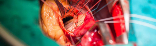
Heart Valve Surgery Heart valve surgery is a procedure to treat heart valve disease. Heart valve disease involves at least one of the four heart valves not working properly. Heart valves keep blood flowing in the correct direction through the heart. The four heart valves are the mitral valve, tricuspid valve, pulmonary valve and aortic valve. Each valve has flaps — called leaflets for the mitral and tricuspid valves and cusps for the aortic and pulmonary valves. These flaps should open and close once during each heartbeat. Valves that don't open or close properly disrupt blood flow through the heart to the body. In heart valve surgery, a surgeon repairs or replaces the damaged or diseased heart valve or valves. Many surgical approaches can be used to repair or replace heart valves, including open-heart surgery or minimally invasive heart surgery. The type of heart valve surgery needed depends on various factors, including your age, your health, and the type and severity of heart valve disease. Robot-assisted heart valve surgery Types Annuloplasty Valvuloplasty Why it's done Heart valve surgery is done to treat heart valve disease. There are two basic types of heart valve problems: A narrowing of a valve (stenosis) A leak in a valve that allows blood to flow backward (regurgitation) You might need heart valve surgery if you have heart valve disease that is affecting your heart's ability to pump blood. If you don't have signs or symptoms, or your condition is mild, your doctor might suggest regular monitoring of the heart valve disease. Lifestyle changes and medications might help manage symptoms. Sometimes, doctors recommend heart valve surgery even for those who don't have symptoms. If you need heart surgery for another condition, doctors might perform heart valve repair or replacement at the same time. Together, you and your doctor should discuss whether heart valve surgery is right for you and if minimally invasive heart surgery is an option. Doctor discusses heart valve disease with a patient. Eventually, your valve might need to be repaired or replaced. In some cases, doctors recommend heart valve repair or replacement even if you're not having symptoms. If you need heart surgery for another condition, doctors might repair or replace the affected heart valve at the same time. Your doctor will discuss with you whether heart valve repair or replacement is more appropriate for your condition. Doctors often recommend heart valve repair when possible, as it preserves your heart valve and might preserve heart function. But sometimes valve replacement is necessary and the best option. Doctors might also evaluate if you're a candidate for minimally invasive heart surgery. Your doctor will discuss the benefits and risks of each procedure. If you need heart valve surgery, if possible, choose a medical center that has performed many heart valve surgeries. Risks Possible heart valve surgery risks include: Bleeding Heart attack Infection Valve dysfunction affecting replaced valves Irregular heart rhythm (arrhythmia) Stroke Death How you prepare Your doctor and treatment team will discuss your heart valve surgery with you and answer any questions. Before being admitted to the hospital for heart valve surgery, talk to your family about your hospital stay and discuss help you'll need when you return home. Food and medications Before you have heart valve surgery, talk to your doctor about: When you can take your regular medications and whether you can take them before your surgery Allergies or reactions you've had to medications When you should stop eating or drinking the night before the surgery Clothing and personal items If you're having heart valve surgery, your treatment team might recommend that you bring several items to the hospital, including: A list of your medications Eyeglasses, hearing aids or dentures Personal care items, such as a brush, a comb, shaving equipment and a toothbrush Loose, comfortable clothing A copy of your advance directive Items that help you relax, such as portable music players or books During heart valve surgery, don't wear: Jewelry Eyeglasses Contact lenses Dentures Nail polish You may need to have your body hair shaved where the incisions will be made. A special soap might be used to wash your skin to help prevent infection. What you can expect during the procedure Incisions in minimally invasive heart surgery and open-heart surgery You'll receive anesthetics to put you in a sleep-like state during the procedure. You'll be connected to a heart-lung bypass machine, which keeps blood moving through your body during the procedure. Heart valve surgery can be performed using standard open-heart surgery, which involves cutting your chest through your breastbone. Minimally invasive heart surgery involves smaller incisions than those used in open-heart surgery. Minimally invasive heart surgery includes surgery performed using long instruments inserted through one or more small incisions in the chest (thoracoscopic surgery), surgery performed through a small incision in the chest, or surgery performed by a surgeon using the assistance of a robot (robot-assisted heart surgery). Minimally invasive heart surgery might involve a shorter hospital stay, quicker recovery and less pain than you'd have with open-heart surgery. Minimally invasive heart surgery ideally should be performed at medical centers with medical teams experienced in performing these types of procedures. Heart valve repair Mitral valve repair Your doctor may often recommend heart valve repair when possible, as it saves the heart valve and may preserve heart function. Heart valve repair surgery may include: Patching holes in a valve Reconnecting valve flaps (leaflets or cusps) Removing excess valve tissue so that the leaflets or cusps can close tightly Replacing cords that support the valve to repair the structural support Separating valve flaps that have fused Tightening or reinforcing the ring around the valve (annulus) Some heart valve repair procedures are done using a long, thin tube (catheter) and clips, plugs or other devices. Doctors might treat a valve with a narrowed valve opening with a catheter procedure called balloon valvuloplasty. A doctor inserts a thin, flexible tube (catheter) with a balloon on the tip into an artery in your arm or groin and guides it to the affected valve. The balloon is inflated, which expands the opening of the heart valve. Doctors then deflate the balloon and remove the catheter and balloon. Heart valve replacement Biological valve replacement Mechanical valve replacement If your heart valve can't be repaired and a catheter-based procedure isn't an option, the valve might need to be replaced. To replace a heart valve, your doctor removes the heart valve and replaces it with a mechanical valve or a valve made from cow, pig or human heart tissue (biological tissue valve). Biological valves often eventually need to be replaced, as they break down over time. If you have a mechanical valve, you'll need to take blood-thinning medications for the rest of your life to prevent blood clots. Doctors will discuss with you the risks and benefits of each type of valve. A minimally invasive catheter procedure might be used to replace certain heart valves. For example, a catheter procedure might be performed to insert a replacement valve into a biological replacement valve in the heart that is no longer working properly. After the procedure After your heart valve surgery, you'll generally spend a day or more in the intensive care unit (ICU). You'll receive fluids and medications through an IV. Other tubes drain urine from the bladder and fluid and blood from the chest. You might be given oxygen through a mask or nasal prongs in your nose. After you complete your stay in the ICU, you'll likely be moved to another hospital room for several days. How long you'll stay in the hospital depends on your condition and surgery. After heart valve surgery, your treatment team will monitor your condition and watch for signs of infection at the incision sites. The team will check your blood pressure, breathing and heart rate. The team will also work with you to manage any pain you have after surgery. You'll likely be asked to walk regularly to gradually increase your activity, and to cough and to do breathing exercises as you recover. You'll be given instructions to follow during your recovery, such as: Watching for signs of infection in your incisions Taking your medications Properly caring for incisions Managing pain and other side effects after your surgery Results After heart valve surgery, your doctor will tell you when you can return to activities. You'll need to attend regular follow-up appointments with your doctor. You might have several tests to evaluate and monitor your condition. Your doctor may recommend making healthy lifestyle changes to keep your heart working well. Examples of heart-healthy lifestyle changes are: Eating a healthy diet Getting regular exercise Managing stress Avoiding tobacco use Source: https://www.mayoclinic.org/tests-procedures/heart-valve-surgery/about/pac-20384901
Implantable Cardioverter-Defibrillators (ICDs)
Heart Procedures
Implantable Cardioverter-Defibrillators (ICDs) An implantable cardioverter-defibrillator (ICD) is a small battery-powered device placed in the chest to detect and stop irregular heartbeats (arrhythmias). An ICD continuously monitors the heartbeat and delivers electric shocks, when needed, to restore a regular heart rhythm. You might need an ICD if you have a dangerously fast heartbeat that keeps your heart from supplying enough blood to the rest of your body (such as ventricular tachycardia or ventricular fibrillation) or if you are at high risk of such a heart rhythm problem (arrhythmia), usually because of a weak heart muscle. An ICD differs from a pacemaker — an implantable device that can prevent dangerously slow heartbeats. Types An ICD is a type of cardiac therapy device. There are two basic types: A traditional ICD is implanted in the chest, and the wires (leads) attach to the heart. The implant procedure requires invasive surgery. A subcutaneous ICD (S-ICD) is another option that's implanted under the skin at the side of the chest below the armpit. It's attached to an electrode that runs along the breastbone. An S-ICD is larger than a traditional ICD but doesn't attach to the heart. Why it's done Subcutaneous implantable cardioverter-defibrillator (S-ICD) An ICD constantly monitors for irregular heartbeats and instantly tries to correct them. It helps when the heart stops beating effectively (cardiac arrest). Your health care provider may recommend an ICD if you've had signs or symptoms of a certain type of irregular heart rhythm called sustained ventricular tachycardia, including fainting. An ICD might also be recommended if you survived a cardiac arrest. Other reasons you may benefit from an ICD are: A history of coronary artery disease and heart attack that has weakened the heart An enlarged heart muscle A genetic heart condition that increases the risk of dangerously fast heart rhythms, such as some types of long QT syndrome Other rare conditions that may affect the heartbeat A health care provider may recommend an S-ICD if there are structural defects in the heart that prevent attaching wires to the heart through the blood vessels. Risks Possible risks of having an ICD implanted include: Infection at the implant site Swelling, bleeding or bruising Blood vessel damage from ICD leads Bleeding around the heart, which can be life-threatening Blood leaking through the heart valve (regurgitation) where the ICD lead is placed Collapsed lung (pneumothorax) Movement (shifting) of the device or leads, which could lead to cardiac perforation (rare) How you prepare Before you get an ICD, your health care provider will order several tests, which may include: Electrocardiography (ECG or EKG). An ECG is a quick and painless test that measures the electrical signals that make the heart beat. Sticky patches (electrodes) are placed on the chest and sometimes the arms and legs. Wires connect the electrodes to a computer, which displays the test results. An ECG can show if the heart is beating too fast, too slow or not at all. Echocardiography. This noninvasive test uses sound waves to create pictures of the heart in motion. It shows the size and structure of the heart and how blood is flowing through the heart. Holter monitoring. A Holter monitor is a small, wearable device that keeps track of the heart rhythm. It may be able to spot irregular heart rhythms that an ECG missed. You typically wear a Holter monitor for 1 to 2 days. Wires from sensors on the chest connect to a battery-operated recording device carried in a pocket or worn on a belt or shoulder strap. While wearing the monitor, you may be asked to keep a diary of your activities and symptoms. Your health care provider will usually compare the diary with the electrical recordings and try to figure out the cause of your symptoms. Event recorder. If you didn't have any irregular heart rhythms while you wore a Holter monitor, your health care provider may recommend an event recorder, which can be worn for a longer time. There are several different types of event recorders. Event recorders are similar to Holter monitors and generally require you to push a button when you feel symptoms. Electrophysiology study (EP study). The health care provider guides a flexible tube (catheter) through a blood vessel into the heart. More than one catheter is often used. Sensors on the tip of each catheter send signals and record the heart's electricity. A health care provider uses this information to identify the area that is causing the irregular heartbeat. What you can expect before the procedure If you're having an ICD implanted, you'll likely be asked to avoid food and drinks for at least 8 hours before the procedure. Talk to your health care provider about any medications you take and whether you should continue to take them before the procedure to implant an ICD. During the procedure A health care provider will insert an IV into your forearm or hand and may give you a medication called a sedative to help you relax. You will likely be given general anesthesia (fully asleep). During surgery to implant the ICD, the doctor guides one or more flexible, insulated wires (leads) into veins near the collarbone to the heart using X-ray images as a guide. The ends of the leads attach to the heart. The other ends attach to a device (shock generator) that's implanted under the skin beneath the collarbone. The procedure to implant an ICD usually takes a few hours. Once the ICD is in place, your doctor will test it and program it for your specific heart rhythm needs. Testing the ICD might require speeding up the heart and then shocking it back into a regular rhythm. Depending on the problem with the heartbeat, an ICD could be programmed for: Low-energy pacing. You may feel nothing or a painless fluttering in your chest when your ICD responds to mild changes in your heartbeat. A higher energy shock. For more-serious heart rhythm problems, the ICD may deliver a higher energy shock. This shock can be painful, possibly making you feel as if you've been kicked in the chest. The pain usually lasts only a second, and there shouldn't be discomfort after the shock ends. Usually, only one shock is needed to restore a regular heartbeat. Some people might have two or more shocks during a 24-hour period. Having three or more shocks in a short amount of time is called an electrical or arrhythmia storm. If you have an electrical storm, you should call 911 or seek emergency medical help to see if your ICD is working properly or if you're having irregular heartbeats. If necessary, the ICD can be adjusted to reduce the number and frequency of shocks. Medications may be needed to make the heart beat regularly and decrease the risk of an ICD electrical storm. After the procedure You'll usually be released on the day after the ICD procedure. You'll need to arrange to have someone to drive you home and help you while you are recovering. The area where the ICD is implanted can be swollen and tender for a few days or weeks. Your health care provider might prescribe pain medication. Aspirin and ibuprofen aren't recommended because they may increase the risk of bleeding. You'll usually need to avoid abrupt movements that raise your left arm above your shoulder for up to eight weeks so the leads don't move until the area has healed. You may need to limit your driving, depending the type of ICD received. Your health care provider will give you instructions on when it's safe to return to driving and other daily activities. For about four weeks after surgery, your health care provider might ask you to avoid: Vigorous, above-the-shoulder activities or exercises, including golf, tennis, swimming, bicycling, bowling or vacuuming Heavy lifting Strenuous exercise programs Your health care provider will probably tell you to avoid contact sports indefinitely. Heavy contact may damage the device or dislodge the wires. Long-term precautions Problems with an ICD due to electrical signals (electrical interference) are rare. Still, take precautions with the following: Cellular phones and other mobile devices. It's safe to talk on a cellphone, but avoid placing a cellphone within 6 inches (about 15 centimeters) of an ICD site when the phone is turned on. Although unlikely, an ICD could mistake a cellphone's signal for a heartbeat and slow the heartbeat, causing symptoms such as sudden fatigue. Security systems. After surgery, you'll receive a card that says you have an ICD. Show your card to airport personnel because the ICD may set off airport security alarms. Also, hand-held metal detectors often contain a magnet that can interfere with an ICD. Limit scanning with a hand-held detector to less than 30 seconds over the site of your ICD or make a request for a manual search. Medical equipment. Tell health care providers that you have an ICD. Some procedures, such as magnetic resonance imaging (MRI), magnetic resonance angiography (MRA), and radiofrequency or microwave ablation may not be recommended if you have an ICD. Your health care provider can tell you if you need an alternative test. Power generators. Stand at least 2 feet (0.6 meters) from welding equipment, high-voltage transformers or motor-generator systems. If you work around such equipment, your health care provider can arrange a test in your workplace to see if the equipment affects your ICD. Headphones and wireless chargers. Headphones may contain a magnetic substance that can interfere with an ICD. Keep headphones and wireless chargers at least 6 inches (about 15 centimeters) from an ICD. Magnets. Magnets may affect an ICD, so it's a good idea to keep magnets at least 6 inches (15 centimeters) from the ICD site. Devices that pose little or no risk to an ICD include microwave ovens, televisions and remote controls, AM/FM radios, toasters, electric blankets, electric shavers and electric drills, computers, scanners, printers, and GPS devices. Driving restrictions If you have an ICD to treat ventricular arrhythmia, driving a vehicle may pose risks to yourself and others. The combination of arrhythmia and shocks from an ICD can cause fainting, which would be dangerous while driving. Many countries have driving restrictions for people with ICDs. If your ICD was implanted due to a previous cardiac arrest or ventricular arrhythmia, your health care provider may recommend waiting several months before driving or operating a vehicle. If you have a shock, with or without fainting, tell your health care provider. Usually, you'll be discouraged from driving until you've been shock-free for several months. If you have an ICD but have no history of life-threatening arrhythmias, you can usually resume driving about a week after your procedure if you've had no shocks. Discuss your situation with your health care provider. You usually can't get a commercial driver's license if you have an ICD. Results An ICD is the main treatment for anyone who has survived cardiac arrest. ICDs are increasingly used in people at high risk of sudden cardiac arrest. An ICD lowers the risk of sudden death from cardiac arrest more than medication alone. Although the electrical shocks can be unsettling, they're a sign that the ICD is effectively treating a heart rhythm problem and protecting against sudden death. The lithium battery in an ICD can last 5 to 7 years. The battery is typically checked during regular checkups, which should occur about every six months. When the battery is nearly out of power, the generator is replaced with a new one during a minor outpatient procedure. ICDs and end-of-life issues If you have an ICD and become terminally ill, your ICD will still deliver shocks if it isn't deactivated. A health care provider can perform a simple procedure to turn off the ICD, if desired. Turning off the device can prevent unwanted shocks and unnecessary suffering. Doing so won't immediately cause the heart to stop. Talk to your health care provider about your wishes. Also talk to family members or the person designated to make medical decisions for you about what you'd like to do in an end-of-life care situation. Source: https://www.mayoclinic.org/tests-procedures/implantable-cardioverter-defibrillators/about/pac-20384692
Minimally Invasive Heart Surgery
Heart Procedures
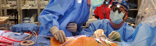
Minimally Invasive Heart Surgery Minimally invasive heart surgery involves making small incisions in the right side of the chest to reach the heart between the ribs, rather than cutting through the breastbone, as is done in open-heart surgery. Minimally invasive heart surgery can be done to treat a variety of heart conditions. Compared with open-heart surgery, this type of surgery might mean less pain and a quicker recovery for many people. Why it's done Many types of heart procedures may be performed with minimally invasive heart surgery, including: Aortic valve replacement Atrial septal defect and patent foramen ovale closure Atrioventricular septal defect surgery Coronary artery bypass surgery Maze procedure for atrial fibrillation Mitral valve repair or replacement Saphenous vein harvest for coronary artery bypass surgery Tricuspid valve repair or replacement The potential benefits of minimally invasive heart surgery when compared with open-heart surgery can include: Less blood loss Lower risk of infection Reduced trauma and pain Shorter time in the hospital, faster recovery and quicker return to normal activities Smaller, less noticeable scars Minimally invasive heart surgery isn't right for everyone. Your doctor and treatment team will work with you to determine whether it's an option to treat your condition. To determine whether minimally invasive heart surgery is the best option, your doctor will likely review your medical history and order tests to get more information about your heart health. Minimally invasive heart surgery is a complex surgical procedure that requires training and experience. You might be referred to a medical center with surgeons and a surgical team who have the needed expertise in performing minimally invasive procedures. Risks Minimally invasive heart surgery can involve risks similar to open-heart surgery, such as: Bleeding Stroke Infection Irregular heart rhythms (arrhythmias) Death Also, it's possible that minimally invasive heart surgery will need to be changed to open-heart surgery if your surgeon thinks it's not safe to continue with the minimal approach. How you prepare Before minimally invasive heart surgery, your doctor and treatment team will explain to you what to expect before, during and after the surgery and potential risks of the surgery. Your doctor and team will discuss concerns you have about your surgery. Your doctor or another member of your treatment team might discuss with you advance directives or other information to consider prior to your surgery. You may need to have your hair shaved at the areas of your body where the procedure will take place. Your skin might be washed with special soap to reduce the risk of infection. Before being admitted to the hospital for your surgery, talk to your family about your hospital stay and discuss help you might need when you return home. Your doctor and treatment team will give you instructions to follow during your recovery when you return home. Food and medications Talk to your doctor about: When you can take your regular medications and whether you can take them before your surgery At what time you should stop eating or drinking the night before the surgery Clothing and personal items Your treatment team might recommend bringing several items to the hospital, including: A list of your medications Eyeglasses, hearing aids or dentures Personal care items, such as a brush, comb, shaving equipment and toothbrush Loose-fitting, comfortable clothing A copy of your advance directive Items that might help you relax, such as a portable music player or books During surgery, avoid wearing: Jewelry Eyeglasses Contact lenses Dentures Nail polish Precautions regarding medications and allergies Talk to your doctor about: Medications you have brought to the hospital and when you should take medications on the day of the procedure Allergies or reactions you have had to medications What you can expect during the procedure Incision sites in robot-assisted heart surgery Minimally invasive heart surgery includes robot-assisted heart surgery, thoracoscopic surgery and surgery through a small incision in the chest (direct less invasive access heart surgery). In all types, surgeons reach your heart through small incisions between the ribs of your chest. A tool with a small video camera inserted through one of the incisions is used to help the surgeon see inside your body. Most minimally invasive procedures use a heart-lung bypass machine, as is used in open-heart surgery. The machine keeps blood moving through your body during the procedure. Robot-assisted heart surgery Robot-assisted heart surgery In robot-assisted heart surgery, the surgeon uses robotic arms, rather than his or her hands, to perform the exact maneuvers used in traditional open-heart surgery. During this procedure, your surgeon works at a remote console and views your heart in a magnified high-definition 3D view on a video monitor. From the console, your surgeon's hand movements translate precisely to the robotic arms at the operating table, which move similarly to the human wrist. A second surgeon and surgical team assist at the operating table, changing surgical instruments attached to the robotic arms. Thoracoscopic surgery In thoracoscopic surgery (sometimes referred to as a minithoracotomy), your surgeon inserts a long, thin tube (thoracoscope) containing a tiny video camera into a small incision in your chest. Your surgeon repairs your heart using long instruments inserted through small incisions between your ribs. After the procedure You'll generally spend a day or so in the intensive care unit (ICU). You'll be given fluids and medications through intravenous (IV) lines. Other tubes placed during surgery will drain urine from your bladder and fluid and blood from your chest. You might be given oxygen through a face mask or prongs in your nose. After the ICU, you'll be moved to a regular hospital room for several days. The time you spend in the ICU and hospital will depend on your condition and surgery. Your treatment team will: Monitor your condition and watch for signs of infection in your incision sites Monitor your blood pressure, breathing and heart rate Work with you to manage pain Get you up and walking and instruct you to gradually increase your activity Show you how to do deep-breathing exercises and instruct you to cough to keep your lungs clear Your doctor will give you instructions to follow during your recovery, such as watching for signs of infection, caring for your incisions, taking medications and managing pain. Your doctor will tell you when you can return to daily activities, such as working, driving and exercise. Results Minimally invasive heart surgery may help reduce aortic stenosis symptoms and improve your quality of life. You'll usually need regular checkups with a doctor to monitor your heart condition. Tests may be done to check your heart health. Your doctor may also recommend following a heart-healthy lifestyle, which includes eating a healthy diet, exercising, managing stress and avoiding tobacco use. Sometimes, a personalized program of education and exercise designed to improve health after heart surgery (cardiac rehabilitation) is also recommended. Source: https://www.mayoclinic.org/tests-procedures/minimally-invasive-heart-surgery/about/pac-20384895
Mitral Valve Repair and Replacement
Heart Procedures
Mitral valve repair and mitral valve replacement are types of surgery to fix or replace a leaky or stiff mitral valve in the heart. The mitral valve is between the left heart chambers (left atrium and left ventricle). Mitral valve repair and mitral valve replacement may be done as an open-heart surgery procedure or as minimally invasive heart surgery. Sometimes a mitral valve problem may be treated with a catheter-based procedure. The specific procedure used depends on the severity of your mitral valve disease and whether it's getting worse. Types Annuloplasty Valvuloplasty Why it's done Surgery or another procedure to repair or replace a mitral valve may be done if you have mitral valve disease such as: Mitral valve regurgitation. The flaps (leaflets) of the mitral valve don't close tightly, causing blood to leak backward. Mitral valve regurgitation is common in people with mitral valve prolapse. Mitral valve repair surgery is recommended if you have severe mitral valve regurgitation symptoms. Mitral valve stenosis. The valve leaflets become thick or stiff, and they may fuse together. This causes the valve to become narrowed and reduces blood flow through the area. If mitral valve disease is mild, your health care provider may first suggest regular checkups to monitor your heart health. You may be prescribed medications to manage symptoms. Sometimes mitral valve repair or mitral valve replacement is done even if you're not having symptoms. Research has found that performing surgery in some people with severe mitral valve regurgitation who aren't having symptoms — rather than monitoring the condition — can improve long-term outcomes. For people with mitral valve disease, health care providers may often recommend repairing the mitral valve when possible, as it preserves the mitral valve and may preserve heart function. Mitral valve repair may be done to avoid complications that can occur with mitral valve replacement, such as blood clots due to mechanical valves and biological tissue valve failure. Risks Possible risks of mitral valve repair and mitral valve replacement surgery include: Bleeding Blood clots Malfunction of the replacement valve Irregular heartbeats (arrhythmias) Infection Stroke Death How you prepare Before mitral valve repair or replacement, an echocardiogram and other tests may be done to get more details about your heart condition. Your care providers will review what to expect during surgery and discuss any concerns you may have. As you prepare for mitral valve repair or replacement, it can be helpful to talk to your loved ones about your hospital stay and what help you may need when you return home. Your care providers will give you specific instructions to follow during your recovery at home. Food and medications Talk to your health care providers about: The medications you take and when or if you can take them before surgery When you should stop eating or drinking the night before the surgery Allergies or reactions you have had to medications Clothing and personal items Your care providers may recommend that you bring several items to the hospital, including: A list of your medications Eyeglasses, hearing aids or dentures Personal care items, such as a brush, a comb, a toothbrush and shaving equipment Loosefitting, comfortable clothing A copy of your advance directive, if you have one Items that may help you relax, such as portable music players or books During surgery, avoid wearing: Contact lenses Dentures Eyeglasses Jewelry Nail polish What you can expect Surgery to repair or replace the mitral valve is done at a hospital by a heart (cardiac) surgeon. If you need heart surgery for another condition in addition to mitral valve disease, the surgeon may perform both surgeries at the same time. Before You'll receive medication to put you into a deep sleep during the procedure (general anesthesia). You'll be connected to a heart-lung bypass machine, which keeps blood moving through your body during the procedure. During Incisions in minimally invasive heart surgery and open-heart surgery Minimally invasive heart surgery Robot-assisted heart surgery Mitral valve repair or replacement may involve: Open-heart surgery, which involves a cut (incision) in the chest. Minimally invasive heart surgery, which uses smaller incisions in the chest. Minimally invasive heart surgery may involve a shorter hospital stay, quicker recovery and less pain than open-heart surgery. Minimally invasive heart surgery generally should be done at medical centers with care providers experienced in performing such surgery. Robot-assisted heart surgery, a type of minimally invasive surgery in which the surgeon uses robotic arms to perform the same movements used in open-heart surgeries. Catheter-based procedure, which is done by a heart doctor in the catheterization lab (interventional cardiologist). This type of treatment involves inserting a thin, flexible tube (catheter) in a vein and guiding it to the heart. The catheter may be used to deliver tools to repair or replace the mitral valve. Mitral valve repair During mitral valve repair surgery, a surgeon may: Patch holes in a heart valve Reconnect the valve leaflets Remove excess tissue from the valve so that the flaps can close tightly Repair the structure of the mitral valve by replacing cords that support it Separate valve leaflets that have fused Other mitral valve repair procedures include: Annuloplasty. Surgeons tighten or reinforce the ring around the valve (annulus). Annuloplasty may be done alone or with other techniques to repair a heart valve. Valvuloplasty. Also called balloon valvotomy, this catheter procedure is used to repair a mitral valve with a narrowed opening. The doctor inserts a catheter with a balloon on the tip into an artery in your arm or groin and guides it to the mitral valve. The balloon is inflated, widening the mitral valve opening. The balloon is deflated, and the catheter and balloon are removed. Mitral valve clip. In this procedure, the surgeon guides a catheter with a clip on its end to the mitral valve through an artery in the groin. The clip is used to fix a torn or leaky mitral valve leaflet. Your health care provider may recommend this option if you have severe mitral valve regurgitation and are not a good candidate for mitral valve surgery. Mitral valve replacement During mitral valve replacement, the heart surgeon removes the mitral valve and replaces it with a mechanical valve or a valve made from cow, pig or human heart tissue (biological tissue valve). Sometimes, a catheter procedure may be done to insert a replacement valve into a biological tissue valve that is no longer working properly. This is called a valve-in-valve procedure. After After mitral valve repair or replacement surgery, you'll generally spend a day or more in the intensive care unit (ICU). How long you'll stay in the ICU and hospital depends on your specific condition and surgery. During your hospital stay, you may receive: Fluids and medications through an IV Oxygen Pain medication Tubes are used to drain urine from the bladder and fluid and blood from the chest. Your care providers will frequently check your blood pressure, breathing and heart rate. After leaving the ICU, you'll usually be moved to a regular hospital room for several days. If you've had a transcatheter procedure, you'll typically stay in the hospital at least overnight but may not need to be in the ICU. With this approach, there are no tubes needed to drain fluid or blood from the chest. As you recover in the hospital you'll be asked to: Gradually increase activity Take longer and longer walks in the hospital Do breathing exercises You'll receive instructions to follow during recovery, which usually includes the following details: How to care for your incisions What medications you should take and when How to manage pain and other side effects The signs and symptoms of infection and when to call your health care provider After mitral valve repair or mitral valve replacement, your health care provider will tell you when you can safely return to daily activities, such as working, driving and exercise. Results Mitral valve repair and replacement surgery may help reduce symptoms and improve quality of life in people with mitral valve disease. If you had mitral valve replacement with a mechanical valve, you'll need blood thinners for life to prevent blood clots. Biological tissue valves break down (degenerate) over time and usually need to be replaced. Regular health checkups are needed to make sure the new or repaired valve is working properly. Your care provider may recommend a program of education and exercise to help you improve your health and recover after heart surgery. This type of program is called cardiac rehabilitation. Following a healthy lifestyle is important to heart health before and after mitral valve surgery. A healthy lifestyle includes: Not smoking Getting regular exercise Eating a healthy diet Managing stress Source: https://www.mayoclinic.org/tests-procedures/mitral-valve-repair-mitral-valve-replacement/about/pac-20384958

Nuclear Stress Test
Heart Procedures
Nuclear Stress Test A nuclear stress test is an imaging test that shows how blood goes to the heart at rest and during exercise. It uses a small amount of radioactive material, called a tracer or radiotracer. The substance is given by IV. An imaging machine takes pictures of how the tracer moves through the heart arteries. This helps find areas of poor blood flow or damage in the heart. A nuclear stress test is one of several types of stress tests. It may be done after a regular exercise stress test, or it may be the first stress test used. If you have coronary artery disease, a nuclear stress test can help your health care provider learn your risk of a heart attack or other heart event. The test uses a positron emission technology (PET) scanner or single-photon emission computed tomography (SPECT) scanner. Other names for a nuclear stress test are: Cardiac PET study. Cardiac SPECT study. Myocardial perfusion imaging (MPI) study. Why it's done This test may be done if you are being treated for heart disease or you have symptoms such as chest pain or shortness of breath. A nuclear stress test is often done to: Diagnose coronary artery disease. The coronary arteries are the major blood vessels that supply the heart with blood, oxygen and nutrients. Coronary artery disease develops when these arteries are damaged or diseased. A nuclear stress test can diagnose coronary artery disease and show how severe the condition is. Create a treatment plan. If you have coronary artery disease, a nuclear stress test can tell your health care provider how well treatment is working. The test also shows how much exercise your heart can handle. This information helps your provider choose the best treatment for you. Source: https://www.mayoclinic.org/tests-procedures/nuclear-stress-test/about/pac-20385231
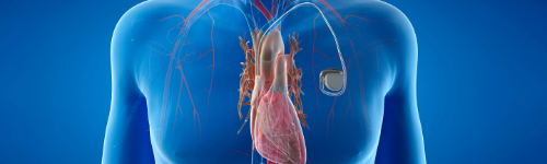
Pacemaker
Heart Procedures
Pacemaker A pacemaker is a small device that's placed (implanted) in the chest to help control the heartbeat. It's used to prevent the heart from beating too slowly. Implanting a pacemaker in the chest requires a surgical procedure. A pacemaker is also called a cardiac pacing device. Types Depending on your condition, you might have one of the following types of pacemakers. Single chamber pacemaker. This type usually carries electrical impulses to the right ventricle of your heart. Dual chamber pacemaker. This type carries electrical impulses to the right ventricle and the right atrium of your heart to help control the timing of contractions between the two chambers. Biventricular pacemaker. Biventricular pacing, also called cardiac resynchronization therapy , is for people who have heart failure and heartbeat problems. This type of pacemaker stimulates both of the lower heart chambers (the right and left ventricles) to make the heart beat more efficiently. Therapy Types Cardiac resynchronization therapy Why it's done A pacemaker is implanted to help control your heartbeat. Your doctor may recommend a temporary pacemaker when you have a slow heartbeat (bradycardia) after a heart attack, surgery or medication overdose but your heartbeat is otherwise expected to recover. A pacemaker may be implanted permanently to correct a chronic slow or irregular heartbeat or to help treat heart failure. The heart's conduction system How your heart beats The heart is a muscular, fist-sized pump with four chambers, two on the left side and two on the right. The upper chambers (right and left atria) and the lower chambers (right and left ventricles) work with your heart's electrical system to keep your heart beating at an appropriate rate — usually 60 to 100 beats a minute for adults at rest. Your heart's electrical system controls your heartbeat, beginning in a group of cells at the top of the heart (sinus node) and spreading to the bottom, causing it to contract and pump blood. Aging, heart muscle damage from a heart attack, some medications and certain genetic conditions can cause an irregular heart rhythm. Pacemaker What a pacemaker does Pacemakers work only when needed. If your heartbeat is too slow (bradycardia), the pacemaker sends electrical signals to your heart to correct the beat. Some newer pacemakers also have sensors that detect body motion or breathing rate and signal the devices to increase heart rate during exercise, as needed. A pacemaker has two parts: Pulse generator. This small metal container houses a battery and the electrical circuitry that controls the rate of electrical pulses sent to the heart. Leads (electrodes). One to three flexible, insulated wires are each placed in one or more chambers of the heart and deliver the electrical pulses to adjust the heart rate. However, some newer pacemakers don't require leads. These devices, called leadless pacemakers, are implanted directly into the heart muscle. Source: https://www.mayoclinic.org/tests-procedures/pacemaker/about/pac-20384689
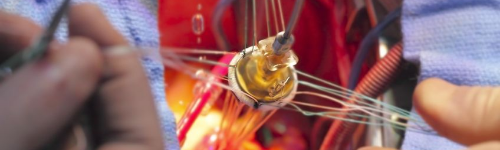
Pulmonary Valve Repair and Replacement
Heart Procedures
Pulmonary Valve Repair and Replacement Pulmonary valve repair and pulmonary valve replacement are procedures to treat a diseased or damaged pulmonary valve. The pulmonary valve is one of four valves that control blood flow in the heart. It's between the lower right heart chamber (right ventricle) and the artery that delivers blood to the lungs (pulmonary artery). A diseased or damaged pulmonary valve can interrupt blood flow from the heart to the lungs. Pulmonary valve repair and pulmonary valve replacement can help improve blood flow and reduce symptoms of heart valve disease. Types Annuloplasty Valvuloplasty Why it's done Pulmonary valve regurgitation Pulmonary valve repair and pulmonary valve replacement are done to treat pulmonary valve disease. Pulmonary valve disease is a condition in which the lung (pulmonary) valve doesn't work properly. It may occur on its own or with other heart conditions present at birth (congenital heart defects), such as tetralogy of Fallot. Types of pulmonary valve disease that may require treatment with pulmonary valve repair or pulmonary valve replacement include: Pulmonary valve regurgitation. A leaky pulmonary valve allows blood to flow backward into the heart (pulmonary valve regurgitation) rather than directly to the lungs for oxygen. Pulmonary valve stenosis. The pulmonary valve is thickened or blocked. It's hard for the valve to open properly. The heart must work harder to pump blood into the pulmonary artery and to the lungs. Pulmonary atresia. This is a congenital heart defect in which a child is born without a well-defined pulmonary valve. In pulmonary atresia, blood can't flow from the right lower heart chamber into the lung artery. The only blood flow to the lungs is through an open passageway between the pulmonary artery and the body's main artery (aorta). The decision to repair or replace a damaged pulmonary valve depends on many things, including: The severity (stage) of pulmonary valve disease Symptoms Age and overall health Whether or not the condition is getting worse Whether surgery is needed to correct another valve or heart condition Pulmonary valve surgery may be done at the same time as other heart valve surgeries. In general, surgeons recommend pulmonary valve repair when possible, as it saves the heart valve and improves heart function. More Information Pulmonary valve repair and replacement care Cardiac MRI Risks All surgeries have risks. Risks of pulmonary valve repair and pulmonary valve replacement depend on your health, the type of procedure, and the expertise of the surgeons and other health care providers. Potential risks of pulmonary valve repair and pulmonary valve replacement include: Bleeding Blood clots Failure of a replacement valve Heart attack Irregular heart rhythms (arrhythmias) Infection Stroke Death How you prepare Pulmonary valve disease consultation Your Mayo Clinic treatment team will discuss any questions you might have about your pulmonary valve repair or pulmonary valve replacement procedure at Mayo Clinic. Before pulmonary valve repair or replacement, your health care providers will explain to you what to expect before, during and after the surgery and potential risks of the surgery. Before being admitted to the hospital for your surgery, talk to your caregivers about your upcoming hospital stay. Discuss any help you may need when you return home. Food and medications Talk to your health care providers about: When or if you can take your regular medications before surgery When or if you should stop eating or drinking the night before surgery Clothing and personal items Your health care providers may recommend that you bring several items to the hospital, including: A copy of your advance directive or living will A list of your medications Eyeglasses, hearing aids or dentures Items that may help you relax, such as portable music players or books Loose-fitting, comfortable clothing Personal care items, such as a brush, comb, shaving equipment and toothbrush Don't wear: Contact lenses Dentures Eyeglasses Jewelry Nail polish What you can expect during the procedure When you arrive for surgery, a care provider will insert an IV into your forearm or hand. Fluids and medicines can be given through the IV. A care provider may shave off any hair at the location where the procedure will take place. For most pulmonary valve repair and replacement procedures, you'll receive medications (general anesthesia) that put you in a sleep-like state so you won't feel any pain during the surgery. You may be connected to a heart-lung bypass machine. The machine keeps blood moving through the body during the heart valve surgery. Pulmonary valve repair Pulmonary valve repair is usually done with open-heart surgery and by opening of the chest bone (sternotomy). Surgeons wire the bone back together after the procedure to prevent movement and aid in healing. Pulmonary valve repair involves: Separating valve flaps that have fused Creating one or more new valve leaflets from your own heart tissues Removing any patches that may have been placed during earlier congenital heart disease surgeries and bringing the pulmonary valve leaflets together to create a working valve Reshaping or removing tissue to allow the valve to close more tightly Tightening or reinforcing the ring around the valve (annulus) Minimally invasive pulmonary valve repair If the pulmonary valve can't fully open due to pulmonary valve stenosis, a less invasive procedure called balloon valvuloplasty may be used to repair the valve. Minimally invasive heart valve surgery typically involves smaller incisions and a shorter hospital stay than traditional open-heart surgery. During balloon valvuloplasty, a surgeon inserts a thin, hollow tube (catheter) into a blood vessel, usually in the groin, and threads it to the heart. A balloon on the tip of the catheter is inflated, which widens the narrowed pulmonary valve. The balloon is deflated and removed. Balloon valvuloplasty is often used to treat infants and children with pulmonary valve stenosis. However, the valve tends to narrow again in adults who have had the procedure. Additional procedures may be needed to treat the narrowed valve over time. Pulmonary valve replacement Pulmonary valve replacement: Biological valve If the pulmonary valve can't be repaired, surgery may be needed to replace the valve. In this procedure, a surgeon removes the pulmonary valve and replaces it with a mechanical valve or a valve made from cow, pig or human heart tissue (biological tissue valve). If you have a mechanical valve, you'll need to take blood-thinning medications for life to prevent blood clots. Biological valves often eventually need to be replaced, as they break down over time. Together, you and your health care providers will discuss the risks and benefits of each type of valve to choose the one that's best for you. Pulmonary valve replacement may be done using open-heart surgery or minimally invasive methods, which involve smaller incisions than those used in open-heart surgery. Minimally invasive pulmonary valve replacement Pulmonary valve-in-valve replacement Minimally invasive pulmonary valve replacement helps reduce the number of open-heart surgeries over a person's lifetime. The most common type of minimally invasive pulmonary valve replacement is transcatheter pulmonary valve replacement, also called percutaneous pulmonary valve replacement. In this type of pulmonary valve replacement, the surgeon inserts a thin, flexible tube (catheter) into a large blood vessel in the groin or chest and guides it to the heart. A replacement pulmonary valve goes through the catheter. A balloon on the tip of the catheter expands to press the new pulmonary valve into place. A transcatheter procedure also may be used to insert a new pulmonary valve in a previously replaced valve that's now failing. This is called a valve-in-valve procedure. After the procedure After open-heart pulmonary valve repair or pulmonary valve replacement, you'll generally spend a day or more in the intensive care unit (ICU). You'll be given fluids, nutrition and medications through intravenous (IV) lines. Other tubes will drain urine from your bladder and drain fluid and blood from your heart and chest. You may be given oxygen. After the ICU, you'll be moved to a regular hospital room for a few days. The time you spend in the ICU and hospital can vary, depending on your condition and procedure. During your hospital stay, your treatment team will: Watch for signs of infection in your incision sites Periodically check your blood pressure, breathing and heart rate Work with you to manage any pain you have after surgery Instruct you to walk regularly to gradually increase your activity and do breathing exercises as you recover You'll receive instructions to follow during your recovery, such as watching for signs of infection in your incisions, properly caring for incisions, taking medications, and managing pain and other side effects after your surgery. Recovery time depends on the specific procedure, your overall health before the procedure and any complications. Results After pulmonary valve repair or pulmonary valve replacement surgery, your health care provider will tell you when you can return to daily activities, such as working, driving and exercise. You'll need regular health checkups to monitor your heart condition and to check for complications, such as valve failure. Imaging tests may be done to make sure the pulmonary valve is working correctly. After heart valve surgery, it's important to follow a heart-healthy lifestyle. Your health care provider may recommend these lifestyle changes: Getting regular exercise Controlling weight Eating a healthy diet Managing stress Not smoking Your care provider may also suggest participating in a program of education and exercise — called cardiac rehabilitation — to improve your overall health and help you recover after pulmonary valve surgery. The results of pulmonary valve repair and replacement depend largely on the skill and expertise of the surgeons and medical center. If you need pulmonary valve repair or replacement, consider being treated at a medical center with a multidisciplinary team of heart surgeons and care providers trained and experienced in heart valve surgery. Source: https://www.mayoclinic.org/tests-procedures/pulmonary-valve-repair-pulmonary-valve-replacement/about/pac-20385090

Pulmonary Vein Isolation
Heart Procedures
Pulmonary Vein Isolation Cardiac ablation Pulmonary vein isolation is a procedure to treat an irregular heart rhythm called atrial fibrillation. Pulmonary vein isolation is a type of cardiac ablation. Cardiac ablation uses heat or cold energy to create tiny scars in the heart to block irregular electrical signals and restore a regular heartbeat. In pulmonary vein isolation, the tiny scars are created in the left upper chamber of the heart in the area where the four lung (pulmonary) veins connect. The pulmonary veins bring oxygen-rich blood from the lungs to the heart. Why it's done Pulmonary vein isolation is done to reduce the signs and symptoms of atrial fibrillation, such as heart palpitations, shortness of breath and weakness. If you have atrial fibrillation, the procedure may help improve your quality of life. Pulmonary vein isolation is usually done after you've tried medications or other treatments first. Risks Possible risks of pulmonary vein isolation include: Bleeding or infection at the site where the catheter was inserted Blood vessel damage Heart valve damage New or worsening heart rhythm problems (arrhythmias) Slow heart rate that could require a pacemaker to correct Blood clots in the legs or lungs (venous thromboembolism) Stroke or heart attack Narrowing of the veins that carry blood between the lungs and heart (pulmonary vein stenosis) Injury or ulceration of the swallowing tube (esophagus), which runs behind the heart Death, rarely Discuss the risks and benefits of cardiac ablation with your health care provider to understand if this procedure is right for you. How you prepare Your health care provider may order several tests to get more information about your heart health before your cardiac ablation. You'll likely need to stop eating and drinking the night before your procedure. Your care provider will tell you how or if you should continue any medications before a cardiac ablation. What you can expect Before Pulmonary vein isolation is done in the hospital. The amount of sedation needed for the procedure depends on the specific arrhythmia and other health conditions. You may be fully awake or lightly sedated, or you may be given general anesthesia (fully asleep). During Catheter insertion points for cardiac ablation The health care provider inserts long, flexible tubes (catheters) through a blood vessel into the heart. The catheters may be inserted through a blood vessel in the groin (most common), shoulder or neck (less common). Depending on the type of ablation, the health care provider may inject dye (contrast) through the catheter, which helps blood vessels show up more clearly on X-ray images. Sensors on the tip of the catheter send electrical impulses and record the heart's electricity. The health care provider uses this information to identify the area that is causing an arrhythmia and to decide where to apply the ablation. This part of the procedure is called an electrophysiology (EP) study. The health care provider moves the catheters from the upper right chamber of the heart to the upper left chamber of the heart where the pulmonary veins connect. Heat (radiofrequency energy) or extreme cold (cryoablation) is used to create small scars in the target area and block the irregular heart rhythms. Usually, each of the four pulmonary veins is treated during pulmonary vein isolation. There may be some minor discomfort when the catheter is moved into the heart and when energy is being delivered. If you have severe pain or shortness of breath during pulmonary vein isolation, let your care provider know. After Pulmonary vein isolation usually takes three to six hours to complete, but may vary depending on the specific arrhythmia. After pulmonary vein isolation, you'll likely be taken to a recovery area to rest quietly for a few hours. A care provider continuously monitors your heartbeat and blood pressure to check for complications of the procedure. Depending on your condition, you may go home the same day or spend the night in the hospital. Plan to have someone else drive you home after your procedure. You may feel a little sore after your procedure, but the soreness shouldn't last more than a week. You'll usually be able to return to your regular activities within a few days after having pulmonary vein isolation. Results Most people see improvements in their quality of life after cardiac ablation, including pulmonary vein isolation. But there's a chance that the irregular heartbeat may return. If this happens, the procedure may be repeated or you and your health care provider might consider other treatments. Pulmonary vein isolation has not been shown to reduce the risk of a stroke, so your health care provider may recommend starting or continuing blood-thinning medications. Source: https://www.mayoclinic.org/tests-procedures/pulmonary-vein-isolation/about/pac-20384996

Supraventricular Tachycardia (SVT) Ablation
Heart Procedures
Supraventricular Tachycardia (SVT) Ablation Supraventricular tachycardia (SVT) ablation uses cold or heat energy to create tiny scars in the heart to block faulty electrical signals and restore the heart rhythm. SVT ablation is used to treat heart rhythm problems (arrhythmias) that affect the heart's upper chambers and cause a fast or erratic heartbeat (supraventricular tachycardia). Why it's done Your health care provider may recommend SVT ablation if you have a condition that causes a fast and erratic heartbeat, such as: Atrial flutter Atrial tachycardia Atrioventricular nodal reentrant tachycardia (AVNRT) Atrioventricular reentrant tachycardia (AVRT) Wolff-Parkinson-White syndrome Orthodromic atrioventricular reciprocating tachycardia Antidromic atrioventricular reciprocating tachycardia Depending on the type of heart rhythm problem, SVT ablation may be one of the first treatments. Sometimes it's done when other medicines or treatments don't work. Together, you and your health care provider can talk about the benefits and risks of SVT ablation. SVT ablation may be an option for children with supraventricular tachycardia who have a high risk of heart-related complications or whose condition hasn't improved with other treatments. What you can expect SVT ablation is done in the hospital. Before the procedure, you'll likely receive a medication called a sedative that helps you relax. A care provider shaves any hair from an area, usually in the groin area, and then numbs the area. The doctor makes a small cut to access a blood vessel in the numbed area, and inserts a long flexible tube (catheter) into the vein. The catheter is gently guided up to the heart. Sensors on the tip of the catheter send electrical impulses and record the heart's electricity. The doctor uses this information to determine the best place to apply the ablation treatment. A doctor will choose one of the following ablation techniques to create small scars in the heart and block irregular heart rhythms. Extreme cold (cryoablation) Heat (radiofrequency energy) Lasers SVT ablation takes about three to six hours. Afterward, you'll be taken to a recovery area where care providers will closely monitor your condition. You'll likely stay overnight in the hospital. Results Most people see improvements in their quality of life after SVT ablation. But there's a chance the fast heartbeat may return. If this happens, the procedure may be repeated or your health care provider might recommend other treatments. Source: https://www.mayoclinic.org/tests-procedures/svt-ablation/pyc-20385010