
Neurological Conditions
All Neurological Conditions

10 Early Signs and Symptoms of Alzheimer's and Dementia
Alzheimers
10 Early Signs and Symptoms of Alzheimer's and Dementia Memory loss that disrupts daily life may be a symptom of Alzheimer's or other dementia. Alzheimer's is a brain disease that causes a slow decline in memory, thinking and reasoning skills. There are 10 warning signs and symptoms. If you notice any of them, don't ignore them. Schedule an appointment with your doctor. The Alzheimer's Association is here to help any time, any day of the year. Call our free 24/7 Helpline at 800.272.3900 to talk to a live person who can answer your questions. Not sure what to ask or where to start? That's okay, too. Just give us a call and we'll guide you from there. 1 - Memory loss that disrupts daily life One of the most common signs of Alzheimer’s disease, especially in the early stage, is forgetting recently learned information. Others include forgetting important dates or events, asking the same questions over and over, and increasingly needing to rely on memory aids (e.g., reminder notes or electronic devices) or family members for things they used to handle on their own. What's a typical age-related change? Sometimes forgetting names or appointments, but remembering them later. 2 - Challenges in planning or solving problems Some people living with changes in their memory due to Alzheimer's or other dementia may experience changes in their ability to develop and follow a plan or work with numbers. They may have trouble following a familiar recipe or keeping track of monthly bills. They may have difficulty concentrating and take much longer to do things than they did before. What's a typical age-related change? Making occasional errors when managing finances or household bills. 3 - Difficulty completing familiar tasks People living with memory changes from Alzheimer's or other dementia often find it hard to complete daily tasks. Sometimes they may have trouble driving to a familiar location, organizing a grocery list or remembering the rules of a favorite game. What's a typical age-related change? Occasionally needing help to use microwave settings or to record a TV show. Talk with a Dementia Expert Now - 24/7 Helpline: 800.272.3900 4 - Confusion with time or place People living with Alzheimer's or other dementia can lose track of dates, seasons and the passage of time. They may have trouble understanding something if it is not happening immediately. Sometimes they may forget where they are or how they got there. What's a typical age-related change? Getting confused about the day of the week but figuring it out later. 5 - Trouble understanding visual images and spatial relationships Some people living with Alzheimer's or other dementia could experience vision changes. This may lead to difficulty with balance or trouble reading. They may also have problems judging distance and determining color or contrast, causing issues with driving. What's a typical age-related change? Vision changes related to cataracts. 6 - New problems with words in speaking or writing People living with Alzheimer's or other dementia may have trouble following or joining a conversation. They may stop in the middle of a conversation and have no idea how to continue or they may repeat themselves. They may struggle with vocabulary, have trouble naming a familiar object or use the wrong name (e.g., calling a "watch" a "hand-clock"). What's a typical age-related change? Sometimes having trouble finding the right word. 7 - Misplacing things and losing the ability to retrace steps A person living with Alzheimer's or other dementia may put things in unusual places. They may lose things and be unable to go back over their steps to find them again. He or she may accuse others of stealing, especially as the disease progresses. What's a typical age-related change? Misplacing things from time to time and retracing steps to find them. 8 - Decreased or poor judgment Individuals living with Alzheimer's or other dementia may experience changes in judgment or decision-making. For example, they may use poor judgment when dealing with money or pay less attention to grooming or keeping themselves clean. What's a typical age-related change? Making a bad decision or mistake once in a while, like neglecting to change the oil in the car. 9 - Withdrawal from work or social activities A person living with Alzheimer’s or other dementia may experience changes in the ability to hold or follow a conversation. As a result, they may withdraw from hobbies, social activities or other engagements. They may have trouble keeping up with a favorite team or activity. What's a typical age-related change? Sometimes feeling uninterested in family or social obligations. 10 - Changes in mood and personality Individuals living with Alzheimer’s or other dementia may experience mood and personality changes. They can become confused, suspicious, depressed, fearful or anxious. They may be easily upset at home, with friends or when out of their comfort zone. What's a typical age-related change? Developing very specific ways of doing things and becoming irritable when a routine is disrupted. When to seek medical advice If you notice one or more signs in yourself or another person, it can be difficult to know what to do. It’s natural to feel uncertain or nervous about discussing these changes with others. Voicing worries about your own health might make them seem more “real.” Or, you may fear upsetting someone by sharing observations about changes in his or her abilities or behavior. However, these are significant health concerns that should be evaluated by a doctor, and it’s important to take action to figure out what’s going on. How is Alzheimer's Disease Diagnosed? Communicating with Your Health Care Team (PDF) 10 Warning Signs Worksheet (PDF) 10 Warning Signs of Alzheimer's (free online course) 10 Steps to Approach Memory Concerns in Others (PDF) 10 Steps to Approach Memory Concerns in Yourself (PDF) Why Get Checked? What is the difference between Alzheimer’s and typical age-related changes? Signs of Alzheimer's and Dementia Typical Age-Related Changes Poor judgment and decision-making Making a bad decision once in a while Inability to manage a budget Missing a monthly payment Losing track of the date or the season Forgetting which day it is and remembering it later Difficulty having a conversation Sometimes forgetting which word to use Misplacing things and being unable to retrace steps to find them Losing things from time to time What to do if you notice these signs If you notice any of the 10 Warning Signs of Alzheimer's in yourself or someone you know, don't ignore them. Schedule an appointment with your doctor. With early detection, you can explore treatments that may provide some relief of symptoms and help you maintain a level of independence longer. Learn About Treatments Prepare for Your Doctor Visit Source: https://www.alz.org/alzheimers-dementia/10_signs

10 Steps to Approach Memory Concerns in Others
Alzheimers
10 Steps to Approach Memory Concerns in Others If you notice changes in friends, family or others close to you and are concerned for their health — particularly when it involves changes in memory, thinking or behavior — it can be difficult to know what to do or say. Although it's natural to be uncertain or nervous about how to offer support, these changes could be a sign of a significant health concern. Use the guide below or print out the PDF to help you feel more confident and prepared as you assess the situation and take action. Don't hesitate to reach out for help — the Alzheimer's Association can connect you with resources and support, including our free 24/7 Helpline (800.272.3900). 1 - What changes in memory, thinking or behavior do you see? What’s the person doing — or not doing — that’s out of the ordinary and causing concern? 2 - What else is going on? Various conditions can cause changes in memory, thinking and behavior. What health or lifestyle issues could be a factor? E.g., family stress or health issues like diabetes or depression. 3 - Learn about the signs of Alzheimer’s and other dementias and the benefits of an early diagnosis. Visit alz.org/10signs to educate yourself on 10 common warning signs of Alzheimer’s and why it’s important to know if dementia is causing the changes. Do you notice any of the signs in the person you’re concerned about? 4 - Has anyone else noticed the change(s)? Find out if friends and family have seen changes. What are they? 5 - Who should have the conversation to discuss concerns? It could be you, a trusted family member or friend, or a combination. It’s usually best to speak one-on-one so that the person doesn’t feel threatened by a group, but use your best judgment to determine what will likely be most comfortable for the individual. 6 - What is the best time and place to have the conversation? Have the conversation as soon as possible. In addition to choosing a date and time, consider where the person will feel most comfortable. 7 - What will you or the person having the conversation say? Try the following: I’ve noticed [change] in you, and I’m concerned. Have you noticed it? Are you worried? How have you been feeling lately? You haven’t seemed like yourself. I noticed you [specific example] and it worried me. Has anything else like that happened? 8 - Offer to go with the person to the doctor. Ask the person if he or she will see a doctor and show your support by offering to go to the appointment. Some words of encouragement may include: There are lots of things that could be causing this, and dementia may or may not be one of them. Let’s see if the doctor can help us figure out what’s going on. The sooner we know what’s causing these problems, the sooner we can address it. I think it would give us both peace of mind if we talked with a doctor. 9 - If needed, have multiple conversations. The first conversation may not be successful. Write down some notes about the experience to help plan for the next conversation. Location Date/time of day What worked well? What didn’t? What was the result? What can be done differently next time? 10 - Turn to the Alzheimer’s Association for information and support. Visit our education resources to take our free Dementia Conversations online program. Learn how to have honest and caring conversations about common concerns — including driving, doctor visits, and legal and financial planning — when someone begins to show signs of dementia. Call our free 24/7 Helpline (800.272.3900) to speak with a master’s-level clinician who can provide more information about how to discuss memory concerns with someone close to you. Visit the Alzheimer’s Association & AARP Community Resource Finder to find local resources, such as health care professionals, and your closest Association chapter. Explore Evaluating Memory and Thinking Problems: What to Expect to learn what a typical medical evaluation may include. Source: https://www.alz.org/alzheimers-dementia/10-steps
Alzheimer's Disease (AD)
Alzheimers
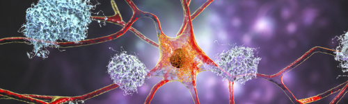
Alzheimer’s Disease (AD) Condition Center — Neurodegenerative Disorders Affecting Memory, Cognition, and Brain Network Integrity Alzheimer’s disease (AD) is the most common cause of dementia worldwide, characterized by progressive decline in memory, executive function, language, and daily functioning. It results from the accumulation of amyloid-beta plaques , tau neurofibrillary tangles , neuroinflammation, and eventual synaptic and neuronal loss—particularly in the hippocampus and association cortices. Although Alzheimer’s is a chronic neurodegenerative disorder, early identification, precision diagnostics (including biomarkers and imaging), and evidence-based treatment strategies can meaningfully slow cognitive decline, improve quality of life, and support patient and caregiver wellbeing. Newly approved disease-modifying therapies targeting amyloid pathology have transformed the treatment landscape. This Condition Center provides a comprehensive framework for recognizing, diagnosing, and managing Alzheimer’s disease with modern standards of care. Questions to Ask Your Doctor • Are my symptoms consistent with Alzheimer’s disease, mild cognitive impairment, or another neurological disorder? • Do I need MRI, PET imaging, or biomarker testing to clarify the diagnosis? • What stage of cognitive impairment am I in, and how will we monitor progression? • Am I a candidate for anti-amyloid monoclonal antibody therapy (e.g., lecanemab, donanemab)? • What non-pharmacologic strategies can support memory, sleep, mood, and brain health? • How should we address driving safety, financial planning, and advance care decisions? • What resources are available for caregiver support and long-term planning? • How do we manage behavioral symptoms, agitation, or sleep disturbances safely? Overview Alzheimer’s disease is a progressive neurodegenerative condition affecting the brain’s memory and cognitive networks. Pathology begins years—sometimes decades—before symptoms appear. Early symptoms typically involve short-term memory deficits, difficulty organizing tasks, word-finding problems, and increased reliance on reminders. As the disease advances, individuals experience greater cognitive impairment, changes in behavior, difficulty completing daily activities, and eventually significant functional dependence. Biological Hallmarks • Amyloid-beta plaques — extracellular protein accumulations • Tau neurofibrillary tangles — intracellular misfolded tau • Synaptic loss and cortical atrophy • Neuroinflammation and microglial activation • Network disconnection—default mode network disruption What Makes Alzheimer’s More Likely? Risk Factors • Age (strongest predictor) • APOE ε4 genotype • Family history of Alzheimer’s • Vascular risk factors (hypertension, diabetes, high cholesterol) • Traumatic brain injury • Low cognitive reserve • Sleep disorders (especially untreated sleep apnea) • Sedentary lifestyle Protective Factors • Higher education/cognitive engagement • Regular exercise • Mediterranean-style diet • Optimal vascular health • Social engagement • Quality sleep Signs and Symptoms Early Stage • Difficulty retaining new information • Repeating questions or stories • Misplacing items • Slowed processing or organization • Word-finding difficulty • Subtle personality changes • Navigation difficulty in unfamiliar places Middle Stage • Difficulty with instrumental activities (finances, medications, cooking) • Impaired judgment • Disorientation to time or place • Language deficits • Behavioral symptoms—irritability, apathy, anxiety • Sleep disturbances • Wandering risk Late Stage • Dependence for basic ADLs • Marked language impairment • Dysphagia • Incontinence • Behavioral disturbances • Increased risk of infections and falls Exams and Tests A thorough diagnostic evaluation helps distinguish Alzheimer’s disease from other dementias (vascular, Lewy body, frontotemporal) and reversible causes of cognitive impairment. 1. Neurological and Cognitive Examination • Montreal Cognitive Assessment (MoCA) • Mini-Mental State Examination (MMSE) • Detailed neuropsychological testing 2. Laboratory Evaluation To rule out reversible contributors: • Vitamin B12 • Thyroid function (TSH, Free T4) • Folate • Liver and kidney function • HIV and syphilis (when clinically indicated) • Thiamine deficiency (if malnutrition or alcohol history) 3. MRI Brain (3 Tesla) Characteristic findings: • Medial temporal lobe and hippocampal atrophy • Parietal lobe volume loss • Enlarged ventricles • Cortical thinning in association cortices • Relative sparing of motor and sensory cortices early Advanced imaging may include: • Volumetric MRI analysis • Diffusion Tensor Imaging (DTI) — white matter tract integrity • SWI — microhemorrhages (important for anti-amyloid therapy eligibility) 4. PET Imaging Amyloid PET Detects amyloid plaque burden. Tau PET Indicates tau tangle distribution—correlates strongly with clinical stage. FDG-PET Hypometabolism in temporoparietal and posterior cingulate regions. 5. CSF Biomarkers Highly reliable indicators of Alzheimer’s pathology: • Low Aβ42 • Elevated total tau • Elevated phosphorylated tau (p-tau) • Aβ42/Aβ40 ratio 6. Blood Biomarkers (rapidly emerging standard) • Plasma p-tau181 / p-tau217 • Neurofilament light chain (NfL) • GFAP (glial fibrillary acidic protein) These are increasingly used for screening and monitoring. Treatment Alzheimer’s disease treatment involves disease-modifying therapy, symptom-targeted interventions, safety planning, and neurowellness strategies that optimize brain health. 1. Disease-Modifying Therapies (Anti-Amyloid Monoclonal Antibodies) Lecanemab (Leqembi) • FDA-approved for early Alzheimer’s disease • Shown to slow cognitive and functional decline • Requires amyloid confirmation via PET or CSF • ARIA (amyloid-related imaging abnormalities) monitoring required Donanemab (anticipated broader approval) • Targets modified forms of amyloid • Produces rapid amyloid clearance • Strong clinical-trial data for slowing cognitive decline Aducanumab (limited use) Not widely used clinically due to reimbursement and efficacy controversies. 2. Symptomatic Therapies Cognitive Symptoms • Cholinesterase inhibitors — donepezil, rivastigmine, galantamine • Memantine — NMDA receptor modulator • Cognitive rehabilitation and structured brain training Behavioral and Psychological Symptoms • Non-pharmacologic strategies first • Treat depression or anxiety with SSRIs when needed • Avoid antipsychotics unless necessary—use lowest effective dose Sleep Disturbances • Treat sleep apnea • Sleep hygiene optimization • Melatonin • Avoid sedatives when possible Movement or Gait Impairment • Physical therapy • Fall-prevention strategies 3. Caregiver and Home Safety • Medication supervision • Financial and legal planning • Driving safety evaluation • Home safety modifications • Wandering precautions (GPS trackers, door alarms) 4. Neurowellness, Longevity & Lifestyle Optimization These strategies directly support brain function and slow decline: Exercise • Aerobic + strength training (≥150 minutes/week) • Improves neuroplasticity and hippocampal volume Nutrition • Mediterranean, MIND, or anti-inflammatory diets • Omega-3 supplementation • Avoid ultra-processed foods Sleep Optimization • Treat apnea • Structured sleep schedule • Reduce nighttime awakenings Cognitive Engagement • Novel learning • Problem-solving activities • Social interaction Vascular Risk Reduction • Control blood pressure, diabetes, cholesterol • Smoking cessation Stress Reduction • Mindfulness • Yoga • Breathing therapy Living With Alzheimer’s Disease Alzheimer’s is a progressive condition, but many individuals can maintain meaningful quality of life for years with early diagnosis, consistent treatment, caregiver support, and lifestyle optimization. Keys to long-term success: • Early identification and staging • Appropriate use of disease-modifying therapies • Sustainable, structured routines • Physical activity • Cognitive stimulation • A strong care network Did You Know? • Alzheimer’s pathology begins up to 20 years before symptoms start. • Amyloid PET and plasma biomarkers allow earlier detection than ever before. • Exercise is the most powerful non-pharmacologic intervention for slowing decline. • ARIA monitoring is essential for patients on anti-amyloid therapies. • Vascular health strongly influences progression speed. References • Diagnostic Criteria & Biomarker Framework (4 references): Jack CR Jr, Bennett DA, Blennow K, et al. NIA-AA Research Framework: Toward a Biological Definition of Alzheimer's Disease. Alzheimers Dement. 2018;14(4):535-562. https://pubmed.ncbi.nlm.nih.gov/29653606 NIA-AA ATN framework: A (amyloid), T (tau), N (neurodegeneration); defines AD biologically using biomarkers regardless of clinical status; amyloid PET or CSF Aβ42/40; tau PET or CSF p-tau; neurodegeneration via MRI or FDG-PET Dubois B, Villain N, Frisoni GB, et al. Clinical Diagnosis of Alzheimer's Disease: Recommendations of the International Working Group. Lancet Neurol. 2021;20(6):484-496. https://pubmed.ncbi.nlm.nih.gov/33946807 IWG-2 criteria: specific cognitive phenotype + in vivo biomarker evidence (amyloid + tau); distinguishes AD from other dementias; emphasizes early detection at MCI stage McKhann GM, Knopman DS, Chertkow H, et al. The Diagnosis of Dementia Due to Alzheimer's Disease: Recommendations From the National Institute on Aging-Alzheimer's Association Workgroups on Diagnostic Guidelines for Alzheimer's Disease. Alzheimers Dement. 2011;7(3):263-269. https://pubmed.ncbi.nlm.nih.gov/21514250 Updated clinical diagnostic criteria: all-cause dementia framework + core AD features (memory-predominant); biomarkers increase diagnostic certainty; staged approach (possible, probable, definite) Hansson O, Edelmayer RM, Boxer AL, et al. The Alzheimer's Association Appropriate Use Recommendations for Blood Biomarkers in Alzheimer's Disease. Alzheimers Dement. 2022;18(12):2669-2686. https://pubmed.ncbi.nlm.nih.gov/36117439 Appropriate use criteria for plasma p-tau217, p-tau181, Aβ42/40; applications in diagnostic workup, clinical trial screening, disease monitoring; sensitivity/specificity >90% for AD pathology Anti-Amyloid Therapies (4 references): van Dyck CH, Swanson CJ, Aisen P, et al. Lecanemab in Early Alzheimer's Disease. N Engl J Med. 2023;388(9):9-21. https://www.nejm.org/doi/full/10.1056/NEJMoa2212948 Clarity AD trial: lecanemab reduced clinical decline by 27% on CDR-SB at 18 months; reduced amyloid PET by 1.21 centiloids; ARIA-E 12.6%, ARIA-H 17.3%; FDA approved for early AD Sims JR, Zimmer JA, Evans CD, et al. Donanemab in Early Symptomatic Alzheimer Disease: The TRAILBLAZER-ALZ 2 Randomized Clinical Trial. JAMA. 2023;330(6):512-527. https://jamanetwork.com/journals/jama/fullarticle/10.1001/jama.2023.13239?utm*source=openevidence&utm*medium=referral TRAILBLAZER-ALZ 2: donanemab slowed decline by 22-35% vs placebo (iADRS); tau-based stratification; 24% achieved amyloid clearance and stopped treatment; ARIA monitoring essential Sevigny J, Chiao P, Bussière T, et al. The Antibody Aducanumab Reduces A β Plaques in Alzheimer's Disease. Nature. 2016;537(7618):50-56. https://pubmed.ncbi.nlm.nih.gov/27582220 Aducanumab phase 1b trial: dose-dependent reduction in amyloid PET; slower decline in MMSE/CDR-SB at highest doses; established proof-of-concept for anti-amyloid immunotherapy Cummings J, Aisen P, Lemere C, et al. Alzheimer's Disease Drug Development Pipeline: 2022. Alzheimers Dement (N Y). 2022;8(1):e12295. https://pubmed.ncbi.nlm.nih.gov/35798474 Pipeline review: 143 agents in clinical trials; 32 in phase 3; anti-amyloid therapies (lecanemab, donanemab, gantenerumab); anti-tau, neuroprotective, metabolic, inflammation-targeting approaches Blood Biomarkers (3 references): Palmqvist S, Janelidze S, Quiroz YT, et al. Discriminative Accuracy of Plasma Phospho-Tau217 for Alzheimer Disease vs Other Neurodegenerative Disorders. JAMA. 2020;324(8):772-781. https://jamanetwork.com/journals/jama/fullarticle/10.1001/jama.2020.12134?utm*source=openevidence&utm*medium=referral Plasma p-tau217 discriminates AD from non-AD with AUC 0.89-0.98; correlates with amyloid and tau PET; detects pathology up to 20 years before symptoms; superior to p-tau181 Hansson O, Lehmann S, Otto M, et al. Advantages and Disadvantages of the Use of the CSF Amyloid β (A β ) 42/40 Ratio in the Diagnosis of Alzheimer's Disease. Alzheimers Res Ther. 2019;11(1):34. https://pubmed.ncbi.nlm.nih.gov/30961663 - CSF Aβ42/40 ratio more accurate than Aβ42 alone; accounts for individual variation in total Aβ production; sensitivity 85-95%, specificity 80-90% for AD pathology 11. Nakamura A, Kaneko N, Villemagne VL, et al. High Performance Plasma Amyloid- β Biomarkers for Alzheimer's Disease. Nature. 2018;554(7691):249-254. https://pubmed.ncbi.nlm.nih.gov/29420472 - Immunoprecipitation-mass spectrometry (IP-MS) measures plasma Aβ42/40; concordance with amyloid PET 90%; potential screening tool before expensive PET imaging Imaging & Pathology (3 references): Jack CR Jr, Wiste HJ, Therneau TM, et al. Associations of Amyloid, Tau, and Neurodegeneration Biomarker Profiles With Rates of Memory Decline Among Individuals Without Dementia. JAMA. 2019;321(23):2316-2325. https://jamanetwork.com/journals/jama/fullarticle/10.1001/jama.2019.7437?utm*source=openevidence&utm*medium=referral - Mayo Clinic Study of Aging: A+T+N+ individuals (all biomarkers abnormal) show steepest memory decline; tau in presence of amyloid drives cognitive decline; staging implications 13. Ossenkoppele R, Schonhaut DR, Schöll M, et al. Tau PET Patterns Mirror Clinical and Neuroanatomical Variability in Alzheimer's Disease. Brain. 2016;139(Pt 5):1551-1567. https://pubmed.ncbi.nlm.nih.gov/26962052 - Tau PET tracer (flortaucipir) shows topographic distribution correlating with clinical phenotypes; temporal predominance in typical AD; posterior cortical atrophy shows occipitoparietal tau 14. Scheltens P, De Strooper B, Kivipelto M, et al. Alzheimer's Disease. Lancet. 2021;397(10284):1577-1590. https://pubmed.ncbi.nlm.nih.gov/33667416 - Comprehensive 2021 review: pathophysiology (amyloid cascade, tau propagation, neuroinflammation), clinical stages, biomarker validation, approved and investigational therapies, prevention strategies Symptomatic Treatments & Interventions (3 references): Schneider LS, Mangialasche F, Andreasen N, et al. Clinical Trials and Late-Stage Drug Development for Alzheimer's Disease: An Appraisal From 1984 to 2014. J Intern Med. 2014;275(3):251-283. https://pubmed.ncbi.nlm.nih.gov/24605808 - Cholinesterase inhibitors (donepezil, rivastigmine, galantamine) show modest symptomatic benefit (2-4 points ADAS-Cog); memantine (NMDA antagonist) benefits moderate-severe AD; combination therapy rationale 16. Livingston G, Huntley J, Sommerlad A, et al. Dementia Prevention, Intervention, and Care: 2020 Report of the Lancet Commission. Lancet. 2020;396(10248):413-446. https://pubmed.ncbi.nlm.nih.gov/32738937 - Lancet Commission: 12 modifiable risk factors account for ~40% of dementia cases; less education, hypertension, hearing loss, smoking, obesity, depression, physical inactivity, diabetes, social isolation, excessive alcohol, TBI, air pollution 17. Ngandu T, Lehtisalo J, Solomon A, et al. A 2 Year Multidomain Intervention of Diet, Exercise, Cognitive Training, and Vascular Risk Monitoring Versus Control to Prevent Cognitive Decline in At-Risk Elderly People (FINGER): A Randomised Controlled Trial. Lancet. 2015;385(9984):2255-2263. https://pubmed.ncbi.nlm.nih.gov/25771249 - FINGER trial: multidomain lifestyle intervention (diet, exercise, cognitive training, vascular monitoring) improved or maintained cognition in at-risk elderly; 25% improvement on neuropsychological test battery vs control Genetics & Risk Factors (3 references): Belloy ME, Napolioni V, Greicius MD. A Quarter Century of APOE and Alzheimer's Disease: Progress to Date and the Path Forward. Neuron. 2019;101(5):820-838. https://pubmed.ncbi.nlm.nih.gov/30790539 - APOE ε4: strongest genetic risk factor for sporadic AD; ε4/ε4 ~10-fold increased risk; influences amyloid deposition, tau pathology, neuroinflammation, lipid metabolism; potential therapeutic target 19. Jansen IE, Savage JE, Watanabe K, et al. Genome-Wide Meta-Analysis Identifies New Loci and Functional Pathways Influencing Alzheimer's Disease Risk. Nat Genet. 2019;51(3):404-413. https://pubmed.ncbi.nlm.nih.gov/30617256 - GWAS meta-analysis: 29 genome-wide significant loci; pathways include immune response, lipid metabolism, tau binding proteins, endocytosis, protein degradation; polygenic risk score development 20. Barnes DE, Yaffe K. The Projected Effect of Risk Factor Reduction on Alzheimer's Disease Prevalence. Lancet Neurol. 2011;10(9):819-828. https://pubmed.ncbi.nlm.nih.gov/21775213 Source: https://www.premierneurohealth.com/
Alzheimer’s Disease (AD)
Neurological Conditions
Alzheimer’s Disease (AD) Condition Center — Neurodegenerative Disorders Affecting Memory, Cognition, and Brain Network Integrity Alzheimer’s disease (AD) is the most common cause of dementia worldwide, characterized by progressive decline in memory, executive function, language, and daily functioning. It results from the accumulation of amyloid-beta plaques , tau neurofibrillary tangles , neuroinflammation, and eventual synaptic and neuronal loss—particularly in the hippocampus and association cortices. Although Alzheimer’s is a chronic neurodegenerative disorder, early identification, precision diagnostics (including biomarkers and imaging), and evidence-based treatment strategies can meaningfully slow cognitive decline, improve quality of life, and support patient and caregiver wellbeing. Newly approved disease-modifying therapies targeting amyloid pathology have transformed the treatment landscape. This Condition Center provides a comprehensive framework for recognizing, diagnosing, and managing Alzheimer’s disease with modern standards of care. Questions to Ask Your Doctor • Are my symptoms consistent with Alzheimer’s disease, mild cognitive impairment, or another neurological disorder? • Do I need MRI, PET imaging, or biomarker testing to clarify the diagnosis? • What stage of cognitive impairment am I in, and how will we monitor progression? • Am I a candidate for anti-amyloid monoclonal antibody therapy (e.g., lecanemab, donanemab)? • What non-pharmacologic strategies can support memory, sleep, mood, and brain health? • How should we address driving safety, financial planning, and advance care decisions? • What resources are available for caregiver support and long-term planning? • How do we manage behavioral symptoms, agitation, or sleep disturbances safely? Overview Alzheimer’s disease is a progressive neurodegenerative condition affecting the brain’s memory and cognitive networks. Pathology begins years—sometimes decades—before symptoms appear. Early symptoms typically involve short-term memory deficits, difficulty organizing tasks, word-finding problems, and increased reliance on reminders. As the disease advances, individuals experience greater cognitive impairment, changes in behavior, difficulty completing daily activities, and eventually significant functional dependence. Biological Hallmarks • Amyloid-beta plaques — extracellular protein accumulations • Tau neurofibrillary tangles — intracellular misfolded tau • Synaptic loss and cortical atrophy • Neuroinflammation and microglial activation • Network disconnection—default mode network disruption What Makes Alzheimer’s More Likely? Risk Factors • Age (strongest predictor) • APOE ε4 genotype • Family history of Alzheimer’s • Vascular risk factors (hypertension, diabetes, high cholesterol) • Traumatic brain injury • Low cognitive reserve • Sleep disorders (especially untreated sleep apnea) • Sedentary lifestyle Protective Factors • Higher education/cognitive engagement • Regular exercise • Mediterranean-style diet • Optimal vascular health • Social engagement • Quality sleep Signs and Symptoms Early Stage • Difficulty retaining new information • Repeating questions or stories • Misplacing items • Slowed processing or organization • Word-finding difficulty • Subtle personality changes • Navigation difficulty in unfamiliar places Middle Stage • Difficulty with instrumental activities (finances, medications, cooking) • Impaired judgment • Disorientation to time or place • Language deficits • Behavioral symptoms—irritability, apathy, anxiety • Sleep disturbances • Wandering risk Late Stage • Dependence for basic ADLs • Marked language impairment • Dysphagia • Incontinence • Behavioral disturbances • Increased risk of infections and falls Exams and Tests A thorough diagnostic evaluation helps distinguish Alzheimer’s disease from other dementias (vascular, Lewy body, frontotemporal) and reversible causes of cognitive impairment. 1. Neurological and Cognitive Examination • Montreal Cognitive Assessment (MoCA) • Mini-Mental State Examination (MMSE) • Detailed neuropsychological testing 2. Laboratory Evaluation To rule out reversible contributors: • Vitamin B12 • Thyroid function (TSH, Free T4) • Folate • Liver and kidney function • HIV and syphilis (when clinically indicated) • Thiamine deficiency (if malnutrition or alcohol history) 3. MRI Brain (3 Tesla) Characteristic findings: • Medial temporal lobe and hippocampal atrophy • Parietal lobe volume loss • Enlarged ventricles • Cortical thinning in association cortices • Relative sparing of motor and sensory cortices early Advanced imaging may include: • Volumetric MRI analysis • Diffusion Tensor Imaging (DTI) — white matter tract integrity • SWI — microhemorrhages (important for anti-amyloid therapy eligibility) 4. PET Imaging Amyloid PET Detects amyloid plaque burden. Tau PET Indicates tau tangle distribution—correlates strongly with clinical stage. FDG-PET Hypometabolism in temporoparietal and posterior cingulate regions. 5. CSF Biomarkers Highly reliable indicators of Alzheimer’s pathology: • Low Aβ42 • Elevated total tau • Elevated phosphorylated tau (p-tau) • Aβ42/Aβ40 ratio 6. Blood Biomarkers (rapidly emerging standard) • Plasma p-tau181 / p-tau217 • Neurofilament light chain (NfL) • GFAP (glial fibrillary acidic protein) These are increasingly used for screening and monitoring. Treatment Alzheimer’s disease treatment involves disease-modifying therapy, symptom-targeted interventions, safety planning, and neurowellness strategies that optimize brain health. 1. Disease-Modifying Therapies (Anti-Amyloid Monoclonal Antibodies) Lecanemab (Leqembi) • FDA-approved for early Alzheimer’s disease • Shown to slow cognitive and functional decline • Requires amyloid confirmation via PET or CSF • ARIA (amyloid-related imaging abnormalities) monitoring required Donanemab (anticipated broader approval) • Targets modified forms of amyloid • Produces rapid amyloid clearance • Strong clinical-trial data for slowing cognitive decline Aducanumab (limited use) Not widely used clinically due to reimbursement and efficacy controversies. 2. Symptomatic Therapies Cognitive Symptoms • Cholinesterase inhibitors — donepezil, rivastigmine, galantamine • Memantine — NMDA receptor modulator • Cognitive rehabilitation and structured brain training Behavioral and Psychological Symptoms • Non-pharmacologic strategies first • Treat depression or anxiety with SSRIs when needed • Avoid antipsychotics unless necessary—use lowest effective dose Sleep Disturbances • Treat sleep apnea • Sleep hygiene optimization • Melatonin • Avoid sedatives when possible Movement or Gait Impairment • Physical therapy • Fall-prevention strategies 3. Caregiver and Home Safety • Medication supervision • Financial and legal planning • Driving safety evaluation • Home safety modifications • Wandering precautions (GPS trackers, door alarms) 4. Neurowellness, Longevity & Lifestyle Optimization These strategies directly support brain function and slow decline: Exercise • Aerobic + strength training (≥150 minutes/week) • Improves neuroplasticity and hippocampal volume Nutrition • Mediterranean, MIND, or anti-inflammatory diets • Omega-3 supplementation • Avoid ultra-processed foods Sleep Optimization • Treat apnea • Structured sleep schedule • Reduce nighttime awakenings Cognitive Engagement • Novel learning • Problem-solving activities • Social interaction Vascular Risk Reduction • Control blood pressure, diabetes, cholesterol • Smoking cessation Stress Reduction • Mindfulness • Yoga • Breathing therapy Living With Alzheimer’s Disease Alzheimer’s is a progressive condition, but many individuals can maintain meaningful quality of life for years with early diagnosis, consistent treatment, caregiver support, and lifestyle optimization. Keys to long-term success: • Early identification and staging • Appropriate use of disease-modifying therapies • Sustainable, structured routines • Physical activity • Cognitive stimulation • A strong care network Did You Know? • Alzheimer’s pathology begins up to 20 years before symptoms start. • Amyloid PET and plasma biomarkers allow earlier detection than ever before. • Exercise is the most powerful non-pharmacologic intervention for slowing decline. • ARIA monitoring is essential for patients on anti-amyloid therapies. • Vascular health strongly influences progression speed. References • Diagnostic Criteria & Biomarker Framework (4 references): Jack CR Jr, Bennett DA, Blennow K, et al. NIA-AA Research Framework: Toward a Biological Definition of Alzheimer's Disease. Alzheimers Dement. 2018;14(4):535-562. https://pubmed.ncbi.nlm.nih.gov/29653606 NIA-AA ATN framework: A (amyloid), T (tau), N (neurodegeneration); defines AD biologically using biomarkers regardless of clinical status; amyloid PET or CSF Aβ42/40; tau PET or CSF p-tau; neurodegeneration via MRI or FDG-PET Dubois B, Villain N, Frisoni GB, et al. Clinical Diagnosis of Alzheimer's Disease: Recommendations of the International Working Group. Lancet Neurol. 2021;20(6):484-496. https://pubmed.ncbi.nlm.nih.gov/33946807 IWG-2 criteria: specific cognitive phenotype + in vivo biomarker evidence (amyloid + tau); distinguishes AD from other dementias; emphasizes early detection at MCI stage McKhann GM, Knopman DS, Chertkow H, et al. The Diagnosis of Dementia Due to Alzheimer's Disease: Recommendations From the National Institute on Aging-Alzheimer's Association Workgroups on Diagnostic Guidelines for Alzheimer's Disease. Alzheimers Dement. 2011;7(3):263-269. https://pubmed.ncbi.nlm.nih.gov/21514250 Updated clinical diagnostic criteria: all-cause dementia framework + core AD features (memory-predominant); biomarkers increase diagnostic certainty; staged approach (possible, probable, definite) Hansson O, Edelmayer RM, Boxer AL, et al. The Alzheimer's Association Appropriate Use Recommendations for Blood Biomarkers in Alzheimer's Disease. Alzheimers Dement. 2022;18(12):2669-2686. https://pubmed.ncbi.nlm.nih.gov/36117439 Appropriate use criteria for plasma p-tau217, p-tau181, Aβ42/40; applications in diagnostic workup, clinical trial screening, disease monitoring; sensitivity/specificity >90% for AD pathology Anti-Amyloid Therapies (4 references): van Dyck CH, Swanson CJ, Aisen P, et al. Lecanemab in Early Alzheimer's Disease. N Engl J Med. 2023;388(9):9-21. https://www.nejm.org/doi/full/10.1056/NEJMoa2212948 Clarity AD trial: lecanemab reduced clinical decline by 27% on CDR-SB at 18 months; reduced amyloid PET by 1.21 centiloids; ARIA-E 12.6%, ARIA-H 17.3%; FDA approved for early AD Sims JR, Zimmer JA, Evans CD, et al. Donanemab in Early Symptomatic Alzheimer Disease: The TRAILBLAZER-ALZ 2 Randomized Clinical Trial. JAMA. 2023;330(6):512-527. https://jamanetwork.com/journals/jama/fullarticle/10.1001/jama.2023.13239?utm*source=openevidence&utm*medium=referral TRAILBLAZER-ALZ 2: donanemab slowed decline by 22-35% vs placebo (iADRS); tau-based stratification; 24% achieved amyloid clearance and stopped treatment; ARIA monitoring essential Sevigny J, Chiao P, Bussière T, et al. The Antibody Aducanumab Reduces A β Plaques in Alzheimer's Disease. Nature. 2016;537(7618):50-56. https://pubmed.ncbi.nlm.nih.gov/27582220 Aducanumab phase 1b trial: dose-dependent reduction in amyloid PET; slower decline in MMSE/CDR-SB at highest doses; established proof-of-concept for anti-amyloid immunotherapy Cummings J, Aisen P, Lemere C, et al. Alzheimer's Disease Drug Development Pipeline: 2022. Alzheimers Dement (N Y). 2022;8(1):e12295. https://pubmed.ncbi.nlm.nih.gov/35798474 Pipeline review: 143 agents in clinical trials; 32 in phase 3; anti-amyloid therapies (lecanemab, donanemab, gantenerumab); anti-tau, neuroprotective, metabolic, inflammation-targeting approaches Blood Biomarkers (3 references): Palmqvist S, Janelidze S, Quiroz YT, et al. Discriminative Accuracy of Plasma Phospho-Tau217 for Alzheimer Disease vs Other Neurodegenerative Disorders. JAMA. 2020;324(8):772-781. https://jamanetwork.com/journals/jama/fullarticle/10.1001/jama.2020.12134?utm*source=openevidence&utm*medium=referral Plasma p-tau217 discriminates AD from non-AD with AUC 0.89-0.98; correlates with amyloid and tau PET; detects pathology up to 20 years before symptoms; superior to p-tau181 Hansson O, Lehmann S, Otto M, et al. Advantages and Disadvantages of the Use of the CSF Amyloid β (A β ) 42/40 Ratio in the Diagnosis of Alzheimer's Disease. Alzheimers Res Ther. 2019;11(1):34. https://pubmed.ncbi.nlm.nih.gov/30961663 - CSF Aβ42/40 ratio more accurate than Aβ42 alone; accounts for individual variation in total Aβ production; sensitivity 85-95%, specificity 80-90% for AD pathology 11. Nakamura A, Kaneko N, Villemagne VL, et al. High Performance Plasma Amyloid- β Biomarkers for Alzheimer's Disease. Nature. 2018;554(7691):249-254. https://pubmed.ncbi.nlm.nih.gov/29420472 - Immunoprecipitation-mass spectrometry (IP-MS) measures plasma Aβ42/40; concordance with amyloid PET 90%; potential screening tool before expensive PET imaging Imaging & Pathology (3 references): Jack CR Jr, Wiste HJ, Therneau TM, et al. Associations of Amyloid, Tau, and Neurodegeneration Biomarker Profiles With Rates of Memory Decline Among Individuals Without Dementia. JAMA. 2019;321(23):2316-2325. https://jamanetwork.com/journals/jama/fullarticle/10.1001/jama.2019.7437?utm*source=openevidence&utm*medium=referral - Mayo Clinic Study of Aging: A+T+N+ individuals (all biomarkers abnormal) show steepest memory decline; tau in presence of amyloid drives cognitive decline; staging implications 13. Ossenkoppele R, Schonhaut DR, Schöll M, et al. Tau PET Patterns Mirror Clinical and Neuroanatomical Variability in Alzheimer's Disease. Brain. 2016;139(Pt 5):1551-1567. https://pubmed.ncbi.nlm.nih.gov/26962052 - Tau PET tracer (flortaucipir) shows topographic distribution correlating with clinical phenotypes; temporal predominance in typical AD; posterior cortical atrophy shows occipitoparietal tau 14. Scheltens P, De Strooper B, Kivipelto M, et al. Alzheimer's Disease. Lancet. 2021;397(10284):1577-1590. https://pubmed.ncbi.nlm.nih.gov/33667416 - Comprehensive 2021 review: pathophysiology (amyloid cascade, tau propagation, neuroinflammation), clinical stages, biomarker validation, approved and investigational therapies, prevention strategies Symptomatic Treatments & Interventions (3 references): Schneider LS, Mangialasche F, Andreasen N, et al. Clinical Trials and Late-Stage Drug Development for Alzheimer's Disease: An Appraisal From 1984 to 2014. J Intern Med. 2014;275(3):251-283. https://pubmed.ncbi.nlm.nih.gov/24605808 - Cholinesterase inhibitors (donepezil, rivastigmine, galantamine) show modest symptomatic benefit (2-4 points ADAS-Cog); memantine (NMDA antagonist) benefits moderate-severe AD; combination therapy rationale 16. Livingston G, Huntley J, Sommerlad A, et al. Dementia Prevention, Intervention, and Care: 2020 Report of the Lancet Commission. Lancet. 2020;396(10248):413-446. https://pubmed.ncbi.nlm.nih.gov/32738937 - Lancet Commission: 12 modifiable risk factors account for ~40% of dementia cases; less education, hypertension, hearing loss, smoking, obesity, depression, physical inactivity, diabetes, social isolation, excessive alcohol, TBI, air pollution 17. Ngandu T, Lehtisalo J, Solomon A, et al. A 2 Year Multidomain Intervention of Diet, Exercise, Cognitive Training, and Vascular Risk Monitoring Versus Control to Prevent Cognitive Decline in At-Risk Elderly People (FINGER): A Randomised Controlled Trial. Lancet. 2015;385(9984):2255-2263. https://pubmed.ncbi.nlm.nih.gov/25771249 - FINGER trial: multidomain lifestyle intervention (diet, exercise, cognitive training, vascular monitoring) improved or maintained cognition in at-risk elderly; 25% improvement on neuropsychological test battery vs control Genetics & Risk Factors (3 references): Belloy ME, Napolioni V, Greicius MD. A Quarter Century of APOE and Alzheimer's Disease: Progress to Date and the Path Forward. Neuron. 2019;101(5):820-838. https://pubmed.ncbi.nlm.nih.gov/30790539 - APOE ε4: strongest genetic risk factor for sporadic AD; ε4/ε4 ~10-fold increased risk; influences amyloid deposition, tau pathology, neuroinflammation, lipid metabolism; potential therapeutic target 19. Jansen IE, Savage JE, Watanabe K, et al. Genome-Wide Meta-Analysis Identifies New Loci and Functional Pathways Influencing Alzheimer's Disease Risk. Nat Genet. 2019;51(3):404-413. https://pubmed.ncbi.nlm.nih.gov/30617256 - GWAS meta-analysis: 29 genome-wide significant loci; pathways include immune response, lipid metabolism, tau binding proteins, endocytosis, protein degradation; polygenic risk score development 20. Barnes DE, Yaffe K. The Projected Effect of Risk Factor Reduction on Alzheimer's Disease Prevalence. Lancet Neurol. 2011;10(9):819-828. https://pubmed.ncbi.nlm.nih.gov/21775213 Source: https://www.premierneurohealth.com/
Amyotrophic Lateral Sclerosis (ALS)
Neurological Conditions
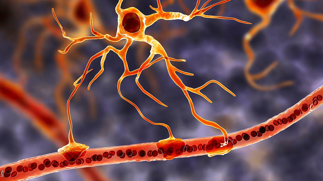
Amyotrophic Lateral Sclerosis (ALS) Your nervous system is the body’s high-speed command network. Motor neurons—specialized nerve cells in the brain and spinal cord—carry signals that let you speak, swallow, breathe, and move. In amyotrophic lateral sclerosis (ALS), also called Lou Gehrig’s disease, these motor neurons gradually stop working and die. As communication between brain and muscle fails, weakness spreads, daily tasks become harder, and—without the right support—breathing can become difficult. ALS is considered a rare disease, yet its impact is anything but rare in the lives it touches. While ALS remains a serious, life-limiting condition, earlier recognition, modern multidisciplinary care, assistive technologies, and several FDA-approved therapies can meaningfully slow decline, extend life, and—crucially—protect quality of life. Many people living with ALS continue to work, parent, create, and advocate for years after diagnosis. Use this condition center to learn what ALS is, how it’s diagnosed and treated, what to expect, and how to partner with your care team. Questions to Ask Your Doctor If you’ve been diagnosed with ALS—or you’re being evaluated for it—bring these questions to your next visit. They’ll help you and your clinicians align on priorities and plan the next steps. What exactly is ALS, and how is it affecting my body right now? Is my ALS limb-onset or bulbar-onset? What does that mean for symptoms and progression? What tests support my diagnosis (EMG, nerve conduction, MRI, labs)? Are there mimicking conditions we should rule out? Should I have genetic testing? If so, which panel, and what would the results change for me or my family? Which medications are appropriate for me now (for example, riluzole, edaravone, or others)? What benefits and side effects should I expect? Am I a candidate for disease-specific therapies such as tofersen (for SOD1-related ALS) or a clinical trial? How do we build my multidisciplinary care team (neurology, respiratory therapy, speech, nutrition, physical/occupational therapy, social work, palliative care)? When should we discuss non-invasive ventilation (BiPAP) and options for communication or mobility devices? What can I do today—exercise, nutrition, sleep, respiratory exercises—to maintain function? How frequently should I have follow-up, and which measures (FVC, SNIP, ALSFRS-R, weight) will we track? Where can I find reliable support groups, home-care resources, and financial/insurance guidance? Overview ALS is a progressive disorder of upper motor neurons (in the brain) and lower motor neurons (in the brainstem and spinal cord). “Amyotrophic” means “no muscle nourishment,” and “lateral sclerosis” refers to scarring on the sides of the spinal cord. As motor neurons fail, muscles weaken and shrink (atrophy). Early symptoms may be subtle—hand clumsiness, foot drop, cramps, or speech changes—and often start in one region before spreading. Although ALS has no cure yet, outcomes are better when care is proactive. Evidence supports: Timely diagnosis and referral to an ALS specialty clinic. Disease-modifying medicines (e.g., riluzole; intravenous or oral edaravone) that can slow progression in selected patients. Targeted therapies for specific genetic forms (e.g., tofersen for SOD1-ALS). Non-invasive ventilation to support breathing, which can prolong survival and improve alertness and comfort. Nutrition optimization , including early gastrostomy when needed to maintain weight and reduce aspiration. Communication and mobility technologies to preserve independence and connection. What Is ALS? ALS is a “motor neuron disease.” Two pathways are affected: Upper motor neurons (brain): damage causes stiffness (spasticity), slowed movement, and brisk reflexes. Lower motor neurons (spinal cord/brainstem): damage causes weakness, cramps, fasciculations (muscle twitches), and muscle wasting. Clinicians often describe the initial pattern: Limb-onset ALS : starts with arm or leg weakness, trouble with tasks like buttoning, writing, or walking. Bulbar-onset ALS : begins with speech/swallowing changes—slurred or nasal speech, coughing when eating, weight loss. Less commonly, breathing may be the first symptom. Cognition is usually preserved early, but up to half of people have mild changes in thinking or behavior, and a smaller group develop a related condition called frontotemporal dementia (FTD) . It’s important to screen for these changes because they influence planning and support. What Makes ALS More Likely? Most ALS is sporadic (no clear family history). About 5–10% is familial , linked to inherited gene variants (e.g., C9orf72 , SOD1 , TARDBP , FUS ). Risk can be influenced by many factors, but having a relative with ALS or FTD is the strongest known predictor. Other observations include: Age : risk rises after age 50. Sex : slightly more common in men before age 65; differences narrow later. Genetics : certain variants increase risk or shape disease features. Environmental/occupational factors : research is ongoing into head trauma, toxins, intense physical activity, and military service; none alone “cause” ALS. Because ALS is complex, risk reduction strategies remain general: protect head/neck, avoid toxins where possible, don’t smoke, and maintain cardiovascular/metabolic health. Early Recognition Matters ALS typically progresses over months to years. Early recognition helps you get ahead of predictable challenges: Safety (falls, aspiration) can be addressed with therapy and home changes. Respiratory support can begin before fatigue and morning headaches worsen. Nutrition can be optimized early to stabilize weight and energy. Clinical trials often require early enrollment. Advance care planning becomes a calm, thoughtful process rather than a crisis decision. Seek urgent evaluation for rapidly advancing weakness , choking/aspiration , or worsening shortness of breath , especially when lying flat or during sleep. Signs and Symptoms Symptoms vary by onset site and rate of progression, but may include: Limb symptoms : hand weakness, dropping objects, tripping, foot drop; muscle cramps and fasciculations. Bulbar symptoms : slurred or nasal speech, trouble projecting voice, choking on liquids/solids, prolonged meals, weight loss. Upper motor neuron signs : stiffness, spasticity, brisk reflexes, clonus. Breathing symptoms : shortness of breath with exertion or at night, restless sleep, morning headaches, daytime sleepiness. Cognitive/behavioral changes : apathy, impulsivity, language difficulties (screened in clinic). Pain is not a direct feature of motor neuron loss, but cramps, spasticity, immobility, and poor posture can cause discomfort—and are treatable. Exams and Tests There’s no single “ALS blood test.” Diagnosis is clinical and supported by testing to confirm motor neuron involvement and rule out mimics. Neurological exam : checks strength, reflexes, tone, speech/swallowing, and gait. Electromyography (EMG) and nerve conduction studies (NCS) : detect active and chronic denervation and reinnervation—electrical footprints of lower motor neuron loss. MRI of brain/spine : excludes structural causes (disc disease, cord compression, stroke, tumor). Laboratory tests : screen for mimics (thyroid, B12, autoimmune, infections, heavy metals). Genetic testing : recommended for familial ALS and increasingly considered in sporadic cases; requires counseling for personal and family implications. Respiratory measures : forced vital capacity (FVC), sniff nasal inspiratory pressure (SNIP), nocturnal oximetry or capnography to guide ventilation timing. Swallowing assessments : speech-language evaluation or videofluoroscopy to tailor nutrition strategies. Treatment While there is no cure yet, care plans combine disease-modifying therapy, symptom management, and supportive technologies. Disease-modifying therapies Riluzole (oral): modestly extends survival by slowing glutamate-mediated toxicity. Edaravone (intravenous or oral): may slow functional decline in selected patients. Tofersen (intrathecal): for SOD1 -related ALS; reduces SOD1 protein and neurofilament biomarkers in many recipients. Your team will discuss benefits, side effects, monitoring, and insurance access. Symptom-targeted care Spasticity/cramps : stretching, physical therapy, baclofen or tizanidine; magnesium, quinine alternatives, or mexiletine may be considered for cramps. Sialorrhea (drooling) : posture, anticholinergic meds, botulinum toxin to salivary glands, or radiotherapy in refractory cases. Speech/communication : early voice banking; low-tech boards to high-tech eye-gaze devices. Swallowing/nutrition : dietary modifications, thickened liquids; feeding tube (PEG) when weight loss or aspiration risk rises—earlier is safer and better tolerated. Breathing support : non-invasive ventilation (BiPAP) at first signs of nocturnal hypoventilation; cough-assist devices and suction help clear secretions. In advanced stages, some choose tracheostomy ventilation; this is a personal decision best discussed early. Mobility and safety : braces, canes, walkers, wheelchairs (including power chairs), home modifications. Mood, sleep, and pain : treat depression/anxiety, optimize sleep, manage pain from immobility. Multidisciplinary clinics (neurology, respiratory therapy, PT/OT, speech, nutrition, social work, mental health, palliative care) improve survival and quality of life. Palliative care focuses on symptom relief and aligning care with your values—from diagnosis onward, not only at end of life. Clinical trials are vital. Ask your team about eligibility for investigational drugs, cell therapies, or biomarker-guided studies. References & Public Resources National Institute of Neurological Disorders and Stroke (NINDS) – ALS Overview Comprehensive summary of ALS causes, symptoms, diagnosis, and current research. Source: https://www.premierneurohealth.com/
Autonomic Disorders, POTS, Dysautonomias, Syncope and Orthostatic Hypotension
Neurological Conditions
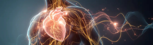
Autonomic Disorders, POTS, Dysautonomias, Syncope and Orthostatic Hypotension The autonomic nervous system controls essential involuntary functions—heart rate, blood pressure, digestion, sweating, temperature regulation, and blood vessel tone. When this system is disrupted, patients experience dizziness, rapid heartbeat, fainting, heat intolerance, fatigue, gastrointestinal symptoms, and significant functional impairment. Autonomic disorders may arise from immune dysfunction, post-viral syndromes, genetic conditions, neuropathies, neurodegenerative diseases, or cardiovascular abnormalities. Postural Orthostatic Tachycardia Syndrome (POTS) and related dysautonomias have become increasingly recognized, particularly following viral infections, including COVID-19. Syncope (fainting) and orthostatic hypotension are among the most common autonomic symptoms and require careful evaluation to rule out cardiac, neurological, or autonomic causes. These conditions are highly impactful but often treatable . With comprehensive autonomic testing and targeted therapy, many individuals regain independence and functionality. Questions to Ask Your Doctor • Are my symptoms due to an autonomic nervous system disorder, cardiovascular issue, or dehydration? • Do I have POTS, neurally mediated syncope, neurogenic orthostatic hypotension, or another form of dysautonomia? • Do I need autonomic testing such as tilt-table testing, heart-rate variability, or sudomotor testing? • Could my condition be autoimmune, post-viral, neuropathic, or neurodegenerative? • Should we evaluate for small fiber neuropathy or post-COVID autonomic dysfunction? • Are medications needed to stabilize heart rate or blood pressure? • What lifestyle strategies can immediately reduce symptoms? • What warning signs require urgent evaluation (chest pain, near-fainting, severe shortness of breath)? • How long does recovery typically take—and can neurowellness strategies improve resilience? Overview This chapter covers four interconnected autonomic disorders: Postural Orthostatic Tachycardia Syndrome (POTS) Dysautonomias (autoimmune, genetic, neuropathic, neurodegenerative, post-viral) Syncope (neurally mediated, autonomic, cardiac) Orthostatic Hypotension (neurogenic and non-neurogenic) All share similar symptoms but have different underlying causes and diagnostic needs. 1. Postural Orthostatic Tachycardia Syndrome (POTS) What It Is A chronic autonomic disorder characterized by: • Heart rate increase ≥30 bpm (≥40 bpm in adolescents) • Occurring within 10 min of standing • Without significant drop in blood pressure Common Symptoms • Lightheadedness • Palpitations • Fatigue • “Brain fog” • Heat intolerance • Exercise intolerance • Nausea • Syncope or near-syncope Common Causes or Associations • Post-viral autonomic dysfunction (including after COVID-19) • Autoimmune disorders (TS-HDS, FGFR3 antibodies) • Small fiber neuropathy • Ehlers-Danlos syndrome • Mast cell activation • Hormonal fluctuations • Deconditioning POTS can be highly disabling but often improves with treatment. 2. Dysautonomias Autonomic dysfunction can result from multiple systemic, autoimmune, genetic, or neurologic causes. Autoimmune Dysautonomia • Associated with ganglionic AChR antibodies • May cause severe orthostatic intolerance, GI dysmotility, anhidrosis Post-Viral Dysautonomia • Frequently seen after SARS-CoV-2 infection • Often overlaps with fatigue, neuropathy, and cognitive symptoms Neuropathic Dysautonomia • Diabetes • Amyloidosis • Chemotherapy-induced neuropathy • Small fiber neuropathy Hereditary Dysautonomia • Familial dysautonomia (HSAN type III) • Mitochondrial disorders Neurodegenerative Dysautonomia Seen in: • Parkinson’s disease • Multiple System Atrophy (MSA) • Lewy body dementia Symptoms include orthostatic hypotension, urinary dysfunction, erectile dysfunction, and constipation. 3. Syncope What It Is Syncope refers to transient loss of consciousness caused by reduced blood flow to the brain. Categories Neurally Mediated (Vasovagal Syncope) • Triggered by standing, pain, emotional stress • Often benign Orthostatic Syncope • Drop in blood pressure upon standing • Can be neurogenic (autonomic failure) or non-neurogenic Cardiac Syncope • Arrhythmias • Structural heart disease • Requires urgent evaluation Symptoms • Pallor • Slow or fast heart rate • Nausea • Lightheadedness • Brief loss of consciousness Red Flags • Occurs during exertion • Occurs without warning • Family history of sudden cardiac death • Associated chest pain or palpitations These require immediate evaluation. 4. Orthostatic Hypotension (OH) What It Is A fall in systolic BP ≥20 mmHg or diastolic BP ≥10 mmHg upon standing. Types Neurogenic OH Due to autonomic failure in: • Parkinson’s disease • Multiple system atrophy • Peripheral neuropathy • Diabetes Non-Neurogenic OH Due to: • Dehydration • Blood loss • Medications (antihypertensives, diuretics) • Cardiovascular disease Symptoms • Dizziness • Blurred vision • Neck heaviness (“coat hanger pain”) • Cognitive slowing • Syncope Signs & Symptoms Across Autonomic Disorders • Dizziness or faintness when standing • Palpitations • Rapid or slow heart rate • Fatigue • Brain fog • Heat intolerance • GI symptoms (nausea, constipation) • Intolerance to standing or exercise • Sweating abnormalities • Anxiety-like symptoms (secondary to physiologic instability) Early Recognition Matters Delayed diagnosis can result in: • Falls and injury from unexpected syncope • Mislabeling physiological symptoms as psychiatric • Missed underlying autoimmune or neuropathic disease • Worsening deconditioning • Reduced quality of life Seek urgent evaluation for: • Syncope during exertion • Suspected arrhythmia • Severe orthostatic hypotension • Chest pain, shortness of breath • New autonomic symptoms after infection or vaccination • Signs of neurodegenerative autonomic failure Exams & Tests Autonomic Function Testing • Tilt-table testing • Heart rate variability • Valsalva maneuver • Deep breathing tests • Quantitative Sudomotor Axon Reflex Test (QSART) • Thermoregulatory sweat testing Laboratory Testing • CBC, CMP • Vitamin B12, methylmalonic acid • Thyroid panel • Autoimmune markers • Ganglionic AChR antibodies • TS-HDS and FGFR3 antibodies (small fiber neuropathy) • Ferritin and iron studies Cardiac Evaluation • ECG • Holter monitor • Echocardiogram • Cardiology referral if red flags present Neuropathy Evaluation • Skin biopsy (small fiber neuropathy) • EMG/NCS for large fiber neuropathy Treatment 1. Lifestyle & Physical Strategies These are foundational for POTS, dysautonomia, and orthostatic intolerance: • Increase fluid intake (2–3 L/day) • Increase salt intake (unless contraindicated) • Compression stockings • Gradual reconditioning and recumbent exercise • Avoid long periods of standing • Elevate head of bed • Frequent small meals 2. Medications Treatment customized based on autonomic subtype: For POTS • Propranolol or ivabradine • Midodrine • Fludrocortisone • Pyridostigmine For Orthostatic Hypotension • Midodrine • Fludrocortisone • Droxidopa • Salt and fluid loading For Dysautonomia • Immunotherapy for autoimmune causes (IVIG, steroids, plasma exchange) • Neuropathic pain medications if small fiber neuropathy present • Management of coexisting mast cell activation or EDS For Syncope • Hydration and salt loading • Counter-pressure maneuvers • Pacemaker in select cardiac cases Neurowellness & Autonomic Longevity Nutritional Optimization • Omega-3 fatty acids • Vitamin D • B-complex vitamins • Hydration-focused diet Anti-Inflammatory & Neuro-Immune Modulation • Curcumin • Magnesium • Sleep restoration Physical Conditioning • Graded exercise therapy (recumbent → upright progression) • Strengthening of core and lower limbs Stress-Response Regulation • Breathwork • Mindfulness • Autonomic retraining exercises Living With Autonomic Disorders Most autonomic disorders improve with: • Accurate diagnosis • Physiologic retraining • Proper medication selection • Lifestyle adjustments • Treatment of underlying conditions Patients often regain independence and functional capacity, although symptoms can fluctuate. Did You Know? • POTS frequently occurs after viral infections due to autonomic nerve injury and immune activation. • Orthostatic hypotension may be an early marker of Parkinson’s disease or multiple system atrophy. • Vasovagal syncope is the most common cause of fainting and is usually benign. • Women are disproportionately affected by POTS and many dysautonomias. • Hydration alone can reduce symptoms by up to 50% in some patients. References (Past 5 Years — Harvard Style) Raj SR, et al. Contemporary understanding and management of POTS. Lancet Neurology. 2020–2024. Vernino S, et al. Autoimmune autonomic ganglionopathy and antibody-mediated dysautonomia. Neurology. 2021–2023. Freeman R, et al. Autonomic disorders and orthostatic intolerance: updated clinical practice. Circulation. 2020–2024. Shouman K, et al. Post-COVID dysautonomia and POTS. JAMA Neurology. 2022–2024. Palma JA, et al. Neurogenic orthostatic hypotension in Parkinson’s and MSA. NEJM. 2020–2023. Sheldon RS, et al. Guidelines for syncope diagnosis and management. Heart Rhythm Society. 2021. Cheshire WP. Autonomic testing and interpretation. Continuum (AAN). 2020–2023. Gibbons CH. Small fiber neuropathy and autonomic dysfunction. Curr Neurol Neurosci Rep. 2021–2024. Kupferberg H, et al. Exercise training in autonomic disorders. Phys Med Rehabil Clin N Am. 2021. Fedorowski A. Autonomic syndromes: pathophysiology and clinical management. Eur Heart J. 2020–2024. Source: https://www.premierneurohealth.com/

Can Alzheimer's Disease Be Prevented?
Alzheimers
Can Alzheimer's Disease Be Prevented? Researchers around the globe are exploring how to prevent Alzheimer's. While Alzheimer's prevention has no definitive answers at this time, research has shown that we can take action to reduce our risk of developing it. What causes Alzheimer's? Prevention studies Heart-head connection Physical exercise and diet Social connections and intellectual activity Head trauma What you can do now Understanding prevention research What causes Alzheimer’s? Experts agree that in the vast majority of cases, Alzheimer's, like other common chronic conditions, probably develops as a result of complex interactions among multiple factors, including age, genetics, environment, lifestyle and coexisting medical conditions. Although some risk factors — such as age or genes — cannot be changed, other risk factors — such as high blood pressure and lack of exercise — usually can be changed to help reduce risk. Research in these areas may lead to new ways to detect those at highest risk. Prevention studies A small percentage of people with Alzheimer’s disease (less than 1%) have an early-onset type associated with genetic mutations. Individuals who have these genetic mutations are guaranteed to develop the disease. An ongoing clinical trial conducted by the Dominantly Inherited Alzheimer Network ( DIAN ), is testing whether antibodies to beta-amyloid can reduce the accumulation of beta-amyloid plaque in the brains of people with such genetic mutations and thereby reduce, delay or prevent Alzheimer's symptoms. Participants in the trial are receiving antibodies (or placebo) before they develop symptoms, and the development of beta-amyloid plaques is being monitored by brain scans and other tests. Though research is still evolving, evidence is strong that people can reduce their risk by making key lifestyle changes, including participating in regular activity and maintaining good heart health. Based on this research, the Alzheimer's Association offers 10 Healthy Habits for Your Brain — a collection of tips that can reduce the risk of cognitive decline. Heart–head connection Several conditions known to increase the risk of cardiovascular disease — such as high blood pressure, diabetes and high cholesterol — also increase the risk of developing Alzheimer's. Some autopsy studies show that as many as 80% of individuals with Alzheimer's disease also have cardiovascular disease. A longstanding question is why some people develop hallmark Alzheimer's plaques and tangles but do not develop the symptoms of Alzheimer's. Vascular disease may help researchers eventually find an answer. Some autopsy studies suggest that plaques and tangles may be present in the brain without causing symptoms of cognitive decline unless the brain also shows evidence of vascular disease. More research is needed to better understand the link between vascular health and Alzheimer’s. Physical exercise and diet Regular physical exercise may be a beneficial strategy to lower the risk of Alzheimer's and vascular dementia. Exercise may directly benefit brain cells by increasing blood and oxygen flow in the brain. Because of its known cardiovascular benefits, a medically approved exercise program is a valuable part of any overall wellness plan. Current evidence suggests that heart-healthy eating may also help protect the brain. Heart-healthy eating includes limiting the intake of sugar and saturated fats and making sure to eat plenty of fruits, vegetables, and whole grains. No one diet is best. Two diets that have been studied and may be beneficial to lowering the risk of Alzheimer's are the DASH (Dietary Approaches to Stop Hypertension) diet and the Mediterranean diet. The DASH diet emphasizes vegetables, fruits, fat-free or low-fat dairy products, whole grains, fish, poultry, beans, seeds, nuts and vegetable oils. The DASH diet limits sodium, sweets, sugary beverages and red meats. A Mediterranean diet includes relatively little red meat. It emphasizes whole grains, fruits and vegetables, fish and shellfish, and healthy fats like nuts and olive oil. Social connections and intellectual activity A number of studies indicate that maintaining strong social connections and keeping mentally active as we age might lower the risk of cognitive decline and Alzheimer's. Experts are not certain about the reason for this association. It may be due to direct mechanisms through which social and mental stimulation strengthen connections between nerve cells in the brain. Head trauma There appears to be a strong link between future risk of cognitive decline and serious head trauma , especially when injury involves loss of consciousness. You can help reduce your risk of Alzheimer's and protect your head by: Wearing a seat belt. Using a helmet when participating in sports. "Fall-proofing" your home by minimizing clutter, loose rugs and poor lighting. What you can do now While research is not yet conclusive — partially due to the need for more large-scale studies in diverse populations — certain lifestyle choices, such as physical activity and diet, may help support brain health and prevent Alzheimer's. Many of these lifestyle changes have been shown to lower the risk of other diseases, like heart disease and diabetes, which have been linked to Alzheimer's. With few drawbacks and plenty of known benefits, healthy lifestyle choices can improve your health and possibly protect your brain. Learn More: Brain Health Participate in clinical research You can help increase our knowledge by considering participation in clinical research. Our free clinical research matching service, TrialMatch®, can help you find clinical trials in your area that are seeking volunteers. Learn More Understanding prevention research Here are some things to keep in mind about the research underlying much of our current knowledge about possible prevention: Insights about potentially modifiable risk factors apply to large population groups, not to individuals. Studies can show that factor X is associated with outcome Y, but cannot guarantee that any specific person will have that outcome. As a result, you can "do everything right" and still have a serious health problem or "do everything wrong" and live to be 100. Much of our current evidence comes from large epidemiological studies such as the Honolulu-Asia Aging Study, the Nurses' Health Study, the Adult Changes in Thought Study and the Kungsholmen Project. These studies explore pre-existing behaviors and use statistical methods to relate those behaviors to health outcomes. This type of study can show an "association" between a factor and an outcome but cannot "prove" cause and effect. This is why we describe evidence based on these studies with such language as "suggests," "may show," "might protect," and "is associated with." The gold standard for showing cause and effect is a clinical trial in which participants are randomly assigned to a prevention or risk management strategy or a control group. Researchers follow the two groups over time to see if their outcomes differ significantly. It is unlikely that some prevention or risk management strategies will ever be tested in randomized trials for ethical or practical reasons. One example is exercise. Definitively testing the impact of exercise on Alzheimer's risk would require a huge trial enrolling thousands of people and following them for many years. The expense and logistics of such a trial would be prohibitive, and it would require some people to go without exercise, a known health benefit. Source: https://www.alz.org/alzheimers-dementia/research-and-progress/prevention
Carpal Tunnel Syndrome (CTS)
Neurological Conditions
Carpal Tunnel Syndrome (CTS) Your hands function as precise communication tools between your brain and the world. Every motion—gripping, typing, lifting, writing—relies on rapid electrical signals traveling through the median nerve , which passes through a narrow passage in the wrist called the carpal tunnel . When this space becomes crowded or its internal pressure increases, the median nerve becomes compressed, disrupting its ability to transmit normal sensation and control fine motor movement. This is known as Carpal Tunnel Syndrome (CTS) , the most common entrapment neuropathy worldwide. CTS may start subtly intermittent tingling, a numb thumb on waking, dropping objects, but, without the right care, it can progress to constant numbness, hand weakness, and even permanent loss of thumb muscle bulk. The good news: CTS is highly treatable. Splints, ergonomic strategies, injections, and—when necessary—surgery can provide lasting relief and prevent further nerve damage. Use this condition center to learn what CTS is, how it’s diagnosed and treated, and how to partner effectively with your care team. Questions to Ask Your Doctor If you’ve been diagnosed with CTS—or are being evaluated for it—these questions can help align you and your healthcare team: • Is my condition definitely CTS, or could my symptoms be coming from my neck, shoulder, or another nerve problem? • How severe is my CTS on examination or nerve testing? Is there evidence of nerve injury? • What factors might be contributing—repetitive activity, ergonomics, diabetes, thyroid issues, inflammation? • Should I start with conservative care such as splinting or therapy? For how long? • Would a corticosteroid injection help confirm the diagnosis or reduce symptoms? • What is the risk of permanent nerve damage if I delay treatment? • When is surgery recommended, and what are the expected outcomes? • How long is recovery, and how soon can I return to work or daily activities? • What ergonomic changes should I make at work or home to prevent recurrence? • How often should I follow up, and what symptoms should prompt earlier reassessment? Overview Carpal Tunnel Syndrome occurs when the median nerve becomes compressed within the rigid walls of the carpal tunnel—a narrow, bony canal in the wrist. Inside this tunnel are nine flexor tendons and the median nerve; any swelling, inflammation, or structural narrowing increases pressure on the nerve. Early on, symptoms are intermittent and often reversible. Over time, however, constant compression can cause progressive sensory loss , motor impairment , and atrophy of the thumb muscles. Although CTS can be frustrating and disruptive, treatment outcomes are excellent when the condition is recognized early and managed proactively. Evidence supports: • Nighttime splinting in a neutral position • Activity and ergonomic modification to reduce repetitive wrist strain • Timely steroid injections for inflammation-driven CTS • Early referral for nerve conduction studies when symptoms persist • Surgical decompression for moderate-to-severe or progressive disease • Addressing systemic conditions such as diabetes, thyroid disease, and arthritis What Is CTS? CTS is a focal entrapment neuropathy of the median nerve at the wrist. As pressure within the carpal tunnel increases, the nerve’s blood supply is reduced, conduction slows, and sensory fibers are the first to malfunction. Over time, motor fibers also weaken, leading to impaired thumb function. Sensory symptoms often begin in the median-innervated digits: • Thumb • Index finger • Middle finger • Radial half of the ring finger Motor symptoms appear later and reflect weakness in thumb abduction and opposition, making it harder to grasp objects, open lids, write, or perform fine motor tasks. Severe or longstanding compression can lead to thenar muscle atrophy , often visible at the base of the thumb. What Makes CTS More Likely? CTS results from multifactorial causes, including: Anatomical or biomechanical factors • Repetitive wrist flexion or extension • Prolonged gripping or vibrating tool use • Wrist fractures, arthritis, or structural narrowing • Tendon sheaths thickening from overuse or inflammation Medical/Systemic conditions • Diabetes • Hypothyroidism • Obesity • Inflammatory arthritis (e.g., rheumatoid arthritis) • Pregnancy-related fluid retention • Chronic kidney disease or dialysis-related amyloidosis Lifestyle and occupational patterns • High-volume keyboard/mouse use • Forceful repetitive manual tasks • Poor wrist posture during work or sleep Although CTS is more common in women and typically arises in midlife, it can affect anyone whose median nerve is exposed to increased tunnel pressure. Early Recognition Matters CTS is a progressive condition. Earlier intervention helps prevent irreversible nerve injury. Recognizing CTS early allows you and your care team to: • Reduce nighttime symptoms with splinting • Modify aggravating activities before structural damage occurs • Perform nerve conduction studies to measure severity • Begin targeted treatment to prevent muscle wasting • Consider surgical decompression before permanent deficits develop Seek urgent evaluation if you notice: • Constant numbness (not intermittent) • Worsening hand weakness • Visible thenar muscle atrophy • Difficulty gripping objects or frequent dropping Signs and Symptoms CTS symptoms vary based on severity and duration but often include: Sensory symptoms • Tingling or numbness in the thumb, index, middle, and part of the ring finger • Nighttime symptoms causing awakening and need to “shake out” the hand • Burning or radiating pain up the forearm • Decreased ability to discriminate shapes, textures, or temperature Motor symptoms • Weak grip or pinch strength • Difficulty opening jars, turning keys, or buttoning clothes • Dropping objects • Thumb clumsiness • Thenar muscle wasting in advanced cases Atypical symptoms suggesting alternate or comorbid pathology: • Pain radiating to the shoulder or neck • Numbness involving the little finger (suggesting ulnar or cervical involvement) Exams and Tests Diagnosis is based on a combination of clinical evaluation and testing: Physical Examination • Tinel’s sign (tapping over the median nerve) • Phalen’s maneuver (wrist flexion to reproduce symptoms) • Durkan’s compression test • Strength assessment of thumb abduction/opposition • Sensory testing of median-innervated digits • Inspection for thenar atrophy Electrodiagnostic Testing (NCS/EMG) The gold standard for diagnosis. Nerve conduction studies evaluate: • Distal latency delay • Slowed conduction velocity across the wrist • Reduced sensory amplitudes EMG identifies: • Chronic median nerve injury • Denervation in thenar muscles • Coexisting radiculopathy or plexopathy Imaging (when indicated) • Ultrasound to assess median nerve swelling • MRI to evaluate structural abnormalities or atypical symptoms Laboratory work may be considered to assess systemic contributors: • Thyroid function • Blood glucose/HbA1c • Vitamin B12 • Inflammatory markers Treatment CTS treatment focuses on relieving pressure on the median nerve, reducing inflammation, restoring function, and preventing permanent injury. 1. Conservative Management Splinting • Neutral wrist splints worn at night • May also be used during daytime repetitive tasks Activity/Ergonomic Modification • Adjust keyboard, mouse, and wrist posture • Reduce repetitive gripping and vibratory tasks • Use supportive wrist rests and proper workstation design Medications • NSAIDs for pain relief • Oral steroids for short-term symptom reduction in selected cases Hand Therapy • Median nerve gliding exercises • Tendon gliding programs • Strengthening and ergonomic training 2. Corticosteroid Injection A local steroid injection into the carpal tunnel can: • Reduce inflammation around the nerve • Provide weeks to months of symptom relief • Aid in diagnosis (improvement strongly supports CTS) Injections are safe and effective but are not a long-term cure for most patients. 3. Surgical Treatment Carpal Tunnel Release Indicated for: • Moderate-to-severe CTS on nerve studies • Progressive weakness or muscle atrophy • Persistent symptoms despite conservative care Both open and endoscopic techniques relieve pressure by dividing the transverse carpal ligament. Success rates exceed 90% when performed in appropriate candidates. Recovery • Nighttime symptoms often improve immediately • Strength returns gradually over weeks to months • Advanced, longstanding nerve damage may be partially reversible Living With CTS With appropriate treatment, most individuals regain normal or near-normal hand function. Maintaining long-term nerve health includes: • Optimizing ergonomics at work and home • Monitoring and managing systemic conditions • Avoiding repetitive strain and extreme wrist positions • Seeking early evaluation if symptoms recur References & Public Resources Palmbergen WAC, Beekman R, Heeren AM, et al. Surgery Versus Corticosteroid Injection for Carpal Tunnel Syndrome (DISTRICTS): An Open-Label, Multicentre, Randomised Controlled Trial. The Lancet . 2025;405(10495):2153-2163. https://pubmed.ncbi.nlm.nih.gov/40517008 Wipperman J, Penny ML. Carpal Tunnel Syndrome: Rapid Evidence Review. American Family Physician . 2024;110(1):52-57. https://www.aafp.org/link_out?pmid=39028782 ** Ashworth NL, Bland JD, Chapman KM, et al. Local Corticosteroid Injection Versus Surgery for Carpal Tunnel Syndrome. The Cochrane Database of Systematic Reviews . 2024;8:CD015101. https://pubmed.ncbi.nlm.nih.gov/39206746 American Academy of Orthopaedic Surgeons. Management of Carpal Tunnel Syndrome: Evidence-Based Clinical Practice Guideline. Published May 2024. https://www.aaos.org/quality/quality-programs/upper-extremity-programs/carpal-tunnel-syndrome/ Yang FA, Wang HY, Kuo TY, et al. Injection Therapy for Carpal Tunnel Syndrome: A Systematic Review and Network Meta-Analysis of Randomized Controlled Trials. PLoS One . 2024;19(5):e0303537. https://pubmed.ncbi.nlm.nih.gov/38753671 Lusa V, Karjalainen TV, Pääkkönen M, et al. Surgical Versus Non-Surgical Treatment for Carpal Tunnel Syndrome. The Cochrane Database of Systematic Reviews . 2024;1:CD001552. https://pubmed.ncbi.nlm.nih.gov/38189479 Padua L, Cuccagna C, Giovannini S, et al. Carpal Tunnel Syndrome: Updated Evidence and New Questions. The Lancet Neurology . 2023;22(3):255-267. https://pubmed.ncbi.nlm.nih.gov/36525982 Ashworth NL, Bland JDP, Chapman KM, et al. Local Corticosteroid Injection Versus Placebo for Carpal Tunnel Syndrome. The Cochrane Database of Systematic Reviews . 2023;2:CD015148. https://pubmed.ncbi.nlm.nih.gov/36722795 Karjalainen TV, Lusa V, Page MJ, et al. Splinting for Carpal Tunnel Syndrome. The Cochrane Database of Systematic Reviews . 2023;2:CD010003. https://pubmed.ncbi.nlm.nih.gov/36848651 Currie KB, Tadisina KK, Mackinnon SE. Common Hand Conditions: A Review. JAMA . 2022;327(24):2434-2445. https://jamanetwork.com/journals/jama/fullarticle/2793415 Source: https://www.premierneurohealth.com/
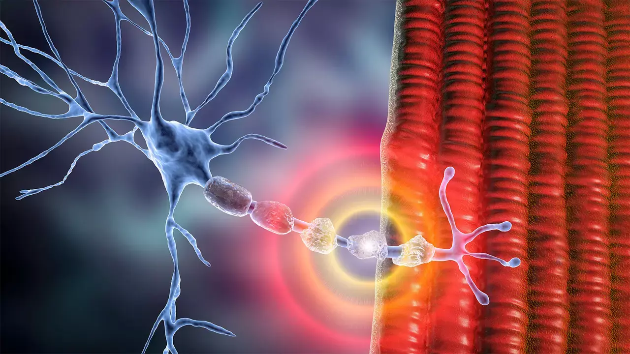
Chronic Inflammatory Demyelinating Polyneuropathy (CIDP)
Neurological Conditions
Chronic Inflammatory Demyelinating Polyneuropathy (CIDP) Your peripheral nerves are the electrical highways connecting your brain and spinal cord to every muscle, joint, and sensation in the body. They send commands that let you walk, talk, feel, and move. When the protective coating on these nerves—called myelin —is damaged by the body’s own immune system, communication slows or stops. This chronic process is known as Chronic Inflammatory Demyelinating Polyneuropathy (CIDP) . CIDP is a long-term, autoimmune neuropathy that causes weakness, numbness, imbalance, and fatigue. Unlike Guillain-Barré Syndrome (GBS) , which develops rapidly over days or weeks, CIDP evolves slowly over months and can fluctuate between relapse and remission. The good news is that CIDP is treatable —and often highly responsive—when recognized early and managed with appropriate therapy. Questions to Ask Your Doctor If you’ve been diagnosed with CIDP or are under evaluation for it, consider asking: What type of CIDP do I have—typical, sensory, multifocal, or motor-predominant? What did my EMG and nerve conduction studies show? Are there antibodies or immune markers that might change my treatment plan? What treatment is best for me right now—IVIG, steroids, plasma exchange, or a newer biologic therapy? How will we track my response to therapy? Are FcRn inhibitors or complement inhibitors available to me? What side effects should I watch for with my medication? Can I switch from IV infusions to a home-based or subcutaneous option? Are there clinical trials or patient registries for CIDP I could join? What physical or lifestyle changes will help protect my nerves long term? Overview CIDP is an autoimmune condition in which the immune system mistakenly attacks myelin in the peripheral nerves. The resulting inflammation and demyelination disrupt signal transmission, causing weakness, sensory loss, and diminished reflexes. Over time, untreated inflammation can lead to secondary axonal injury—permanent nerve fiber damage. CIDP is classified as an acquired immune-mediated demyelinating neuropathy , not an inherited one. It can affect anyone, at any age, and is slightly more common in men. Prevalence is estimated between 1 and 8 per 100,000 people , making it rare—but it remains one of the most treatable chronic neuropathies when detected early. Causes and Mechanism CIDP arises when immune cells—especially T cells, macrophages, and antibodies—mistakenly identify myelin proteins as foreign. This triggers inflammation, complement activation, and segmental demyelination along peripheral nerves. Some patients develop antibodies against specific “nodal” proteins (e.g., neurofascin-155, contactin-1, CASPR1), which may define unique CIDP subtypes and responses to treatment. Researchers continue to study environmental and genetic triggers, but most cases appear idiopathic —without a single clear cause. Symptoms CIDP typically develops slowly over at least 8 weeks and can present in symmetrical or patchy patterns. Common symptoms include: Gradual muscle weakness in arms and legs Numbness , tingling, or burning in hands and feet Loss of reflexes Balance problems and unsteady gait Fatigue or heaviness in limbs Pain or hypersensitivity to touch In rare cases, cranial nerve involvement leading to facial weakness or swallowing difficulty Because CIDP shares features with other conditions (such as diabetic neuropathy or spinal stenosis), precise diagnosis by a neuromuscular specialist is essential. Diagnosis and Tests CIDP is diagnosed through a combination of clinical examination, electrophysiological studies, and supportive laboratory tests. Key tools include: Electromyography (EMG) and Nerve Conduction Studies (NCS): reveal demyelination through slowed conduction velocity, conduction block, or prolonged distal latency. Lumbar Puncture: often shows elevated protein with normal white cell count (albuminocytologic dissociation). MRI of nerve roots or plexus: can show nerve enlargement or enhancement due to inflammation. Blood tests: check for diabetes, thyroid disease, vitamin deficiency, paraproteins, and autoimmune markers. Antibody testing: identifies specific nodal/paranodal antibodies that may guide targeted therapy. Nerve biopsy: occasionally performed to confirm demyelination and rule out vasculitic or infiltrative neuropathies. Treatment The central goal of CIDP treatment is to halt immune attack , allow myelin repair , and restore strength and function . The last few years have transformed therapy—from broad immune suppression to targeted precision biologics that address specific immune pathways. 1. First-Line Immunotherapy These are standard and remain highly effective for most patients: Intravenous Immunoglobulin (IVIG): pooled antibodies that neutralize harmful immune activity. Typically leads to improvement within 2–6 weeks. Subcutaneous Immunoglobulin (SCIG): a home-administered alternative used for maintenance once stability is achieved. It provides steady immunoglobulin levels with fewer infusion side effects. Corticosteroids: such as prednisone or high-dose methylprednisolone, suppress inflammation. They are effective but require monitoring for long-term side effects (weight gain, diabetes, osteoporosis). Plasma Exchange (Plasmapheresis): removes circulating antibodies directly from the bloodstream. It works rapidly and can be lifesaving in severe or refractory cases. Most patients respond to one of these within weeks, and combination therapy may be used to achieve stability before transitioning to long-term maintenance. 2. Advanced and Emerging Therapies New therapies are reshaping the landscape of CIDP care. These treatments aim to modulate specific immune pathways rather than suppress the entire immune system. a. FcRn Inhibitors (Neonatal Fc Receptor Blockade) The neonatal Fc receptor (FcRn) helps recycle IgG antibodies, prolonging their half-life. Blocking FcRn causes harmful antibodies to be broken down faster, reducing inflammation at the nerve. Efgartigimod alfa (Vyvgart Hytrulo) was FDA-approved in 2024 for adults with CIDP after demonstrating reduced relapse risk in the ADHERE Phase III trial. Other investigational FcRn inhibitors— nipocalimab , rozanolixizumab , and batoclimab —are in advanced clinical trials. These agents act within days, are well tolerated, and offer a new option for patients who relapse on IVIG or wish to avoid infusions. b. Complement Inhibitors In some forms of CIDP, complement proteins—part of the body’s immune defense—damage the myelin sheath. Complement inhibitors block this cascade at its origin. Riliprubart , a C1s inhibitor, and ravulizumab , a C5 blocker, are under investigation for antibody-mediated CIDP. These biologics have already changed outcomes in diseases like myasthenia gravis and hold promise for treatment-resistant CIDP. Complement inhibition represents the next frontier of precision immunotherapy , especially for nodal/paranodal antibody-positive patients. c. B-cell and Plasma-cell Depletion Certain CIDP variants involve long-lived B-cells and plasma cells that continuously produce damaging antibodies. Rituximab (anti-CD20) is used off-label in refractory CIDP, particularly for those with anti-neurofascin or contactin antibodies. Obinutuzumab and daratumumab (anti-CD38) are being studied as next-generation agents to target resistant antibody-producing cells more effectively. These drugs can induce remission but require careful infection monitoring and specialist oversight. d. Cellular and Regenerative Therapies Experimental research is exploring immune “reset” and nerve repair through cell-based approaches: Autologous Hematopoietic Stem Cell Transplantation (HSCT): reboots the immune system, potentially inducing long-term remission in severe, treatment-resistant cases. Mesenchymal Stem Cell Therapy: may modulate inflammation and support nerve regeneration, though still in clinical trials. Regenerative medicine and Schwann-cell replacement therapies aim to promote remyelination after inflammation resolves. These strategies remain investigational but point to a future where nerve repair, not just immune suppression, becomes central to care. Maintenance and Long-Term Management Once symptoms are controlled, treatment focuses on maintaining remission and minimizing relapse. SCIG is often favored for long-term therapy due to convenience and stable antibody levels. Periodic reassessment ensures patients are not over- or undertreated; many can taper therapy over time. Physical and occupational therapy preserve muscle tone and balance. Pain management may include gabapentin, pregabalin, duloxetine, or topical lidocaine. Lifestyle measures —adequate rest, nutrition, hydration, and avoiding overexertion—help sustain nerve recovery. Research Horizons CIDP treatment is moving rapidly toward individualized, mechanism-based medicine. Areas of active research include: Biomarker-driven therapy: tailoring biologics to antibody status and complement activation. Digital and AI-driven monitoring: wearable devices that track gait and strength changes. Neuroregenerative therapeutics: cell-based and gene-editing technologies to promote remyelination. Combination biologics: FcRn plus complement inhibition to provide deeper, sustained disease control. For many patients, the outlook has never been brighter. Early recognition and multidisciplinary management can now lead not only to remission, but to meaningful functional recovery. Living With CIDP Most people with CIDP can live active, fulfilling lives. Regular follow-up, physical therapy, and strong communication with the care team are vital. Emotional support—from patient groups or counseling—can ease uncertainty and isolation. Practical strategies include pacing activities, using braces or assistive devices when needed, and celebrating small improvements as signs of nerve repair. Did You Know? CIDP is the most treatable chronic neuropathy and often reversible if addressed early. FcRn inhibitors are the first new class of CIDP drugs approved in over 30 years. Some CIDP variants do not respond well to IVIG but improve dramatically with targeted biologics. Exercise, good sleep, and nutrition support nerve health and recovery. References & Public Resources NINDS – Chronic Inflammatory Demyelinating Polyneuropathy (CIDP) https://www.ninds.nih.gov/health-information/disorders/chronic-inflammatory-demyelinating-polyneuropathy-cidp GARD (NIH) – CIDP Overview https://rarediseases.info.nih.gov/diseases/6668/chronic-inflammatory-demyelinating-polyneuropathy Muscular Dystrophy Association – CIDP Resource Page https://www.mda.org/disease/chronic-inflammatory-demyelinating-polyneuropathy American Academy of Neurology (AAN) CIDP Guideline Summary (2021) https://www.aan.com/Guidelines/home/GetGuidelineContent/956 FDA – Approval of Efgartigimod Alfa (Vyvgart Hytrulo) for CIDP (2024) https://www.fda.gov/news-events/press-announcements/fda-approves-efgartigimod-alfa-and-hyaluronidase-cidp Sanofi Press Release – Riliprubart Complement Inhibitor Trial in CIDP (2024) https://www.sanofi.com/en/media-room/press-releases/2024/2024-06-25-20-30-00-2904145 BMJ JNNP – “Evolving Immunopathogenesis of CIDP” (2024 Review) https://jnnp.bmj.com/content/early/2024/10/01/jnnp-2024-334165.full.pdf Hematology Advisor – Stem Cell Therapy in CIDP https://www.hematologyadvisor.com/news/stem-cell-hsct-cidp-polyradiculoneuropathy-patients-benefit-treatment/ ClinicalTrials.gov – CIDP Active Studies https://clinicaltrials.gov/search?cond=chronic%20inflammatory%20demyelinating%20polyneuropathy Source: https://www.premierneurohealth.com/
Cluster Headache, Trigeminal Autonomic Cephalalgias (TACs) and Tension-Type Headache
Neurological Conditions
Cluster Headache, Trigeminal Autonomic Cephalalgias (TACs) and Tension-Type Headache Headache disorders range from the relatively common and mild to some of the most severe pain conditions known in medicine. This Condition Center integrates two major categories: Cluster Headache & Trigeminal Autonomic Cephalalgias (TACs) – Rare, severe, and highly disabling unilateral headache syndromes with autonomic symptoms. Tension-Type Headache (TTH) – The most common primary headache disorder, often driven by stress, neck tension, and musculoskeletal factors. Although distinct in mechanism and presentation, both conditions significantly affect quality of life and require accurate diagnosis, personalized therapy, and lifestyle-based interventions. This unified resource outlines how to recognize, evaluate, and treat these conditions within a structured, neurology-driven framework. Questions to Ask Your Doctor For Cluster Headache & TACs • Do my symptoms fit cluster headache, paroxysmal hemicrania, SUNCT/SUNA, or another TAC? • Do I need MRI imaging to rule out secondary causes such as pituitary or cavernous sinus lesions? • Should I try high-flow oxygen or rapid-acting medications like sumatriptan? • Which preventive options are best for me—verapamil, lithium, CGRP inhibitors, melatonin? • Would an indomethacin trial clarify my diagnosis? • Am I a candidate for nerve blocks or neuromodulation? For Tension-Type Headache • Are my headaches related to posture, stress, or muscle tension? • Should I start preventive therapy or rely on non-medication approaches? • Would physical therapy or TMJ evaluation help reduce symptoms? • How do I prevent medication-overuse headaches? • Are sleep or ergonomic issues contributing? Overview Cluster headache and TACs represent severe neurovascular disorders involving hypothalamic dysfunction, trigeminal activation, and parasympathetic autonomic pathways. They are among the most painful conditions in neurology. Tension-type headache, in contrast, reflects musculoskeletal tension, central pain processing, and biopsychosocial stress , and is vastly more common. These two headache categories are considered together because: • They frequently coexist with migraine and other primary headache disorders • They require exclusion of secondary causes • They respond to structured care pathways • They benefit from lifestyle, ergonomic, and neuromodulatory interventions • Patients often struggle to differentiate them without clinical guidance Together, these disorders form a major portion of headache-related disability in neurology. What Are Cluster Headache & TACs? TACs include: • Cluster headache • Paroxysmal hemicrania • Hemicrania continua • SUNCT/SUNA syndromes All share: • Severe unilateral head pain • Ipsilateral autonomic symptoms (tearing, nasal congestion, facial sweating, eye redness) • Agitation or restlessness during attacks • Circadian or seasonal patterns (particularly cluster headache) Cluster Headache Characterized by: • Explosive, severe orbital/temporal pain • Attacks lasting 15–180 minutes • Occurring in “clusters” for weeks or months • Often triggered by alcohol during active cycles • Nighttime attacks linked to hypothalamic rhythms Paroxysmal Hemicrania / Hemicrania Continua Defined by complete responsiveness to indomethacin , making this medication essential for diagnosis. SUNCT/SUNA Short-lasting attacks (seconds) with dramatic redness and tearing. What Is Tension-Type Headache (TTH)? TTH involves: • Bilateral pressure or “band-like” tightness • Pericranial muscle tenderness • Stress-related exacerbation • Rare or minimal nausea and sensory hypersensitivity • No autonomic symptoms TTH is categorized as: • Episodic (infrequent or frequent) • Chronic TTH (≥15 days/month) Many individuals have mixed migraine + TTH patterns. What Makes These Conditions More Likely? Cluster/TACs • Male sex (particularly cluster headache) • Smoking history • Circadian rhythm disruption • Family history • Hypothalamic abnormalities Tension-Type Headache • Stress, anxiety, sleep disruption • Prolonged computer/desk work • Neck and shoulder strain • TMJ dysfunction or bruxism • Dehydration • Eye strain • Posture imbalance Signs and Symptoms Cluster Headache & TACs • Severe unilateral orbital/temporal pain • Tearing, eye redness • Nasal congestion or runny nose • Eyelid droop or swelling • Facial sweating • Restlessness or pacing • Stereotyped attacks at the same time each day Paroxysmal Hemicrania • Short attacks (2–30 minutes) • ≥5 attacks/day • Rapid, complete response to indomethacin Hemicrania Continua • Continuous unilateral headache with periods of severe exacerbation • Indomethacin-responsive SUNCT/SUNA • Very brief attacks (seconds) • Prominent autonomic symptoms Tension-Type Headache • Bilateral pressure or squeezing sensation • Tightness in forehead, temples, or neck • Mild–moderate intensity • No nausea or vomiting • Worsens with stress, prolonged sitting, or poor posture • Improves with rest or relaxation Exams and Tests 1. MRI Brain with and without Contrast Essential in TACs to exclude: • Pituitary adenoma • Cavernous sinus lesions • Posterior fossa abnormalities MRI is optional in classic TTH but recommended if atypical features exist. 2. Indomethacin Test Diagnostic for: • Paroxysmal hemicrania • Hemicrania continua 3. Musculoskeletal / Cervical Evaluation Important for TTH: • Neck mobility • Scapular and paraspinal tension • Posture analysis • Ergonomic risk factors 4. TMJ and Dental Assessment Bruxism and jaw clenching often drive chronic TTH. 5. Sleep Evaluation Circadian disturbance contributes to cluster cycles and chronic TTH. Treatment A. Treating Cluster Headache & TACs Acute Treatment • High-flow oxygen (12–15 L/min) — most effective non-pharmacologic therapy • Subcutaneous sumatriptan • Intranasal zolmitriptan • Short steroid taper to break cluster cycles For SUNCT/SUNA: • IV lidocaine for rapid control For hemicrania syndromes: • Indomethacin is both diagnostic and therapeutic Preventive Treatment Cluster headache: • Verapamil — gold standard • Galcanezumab (CGRP mAb) — approved for episodic cluster • Lithium • Topiramate • Melatonin • Occipital nerve block SUNCT/SUNA: • Lamotrigine • Topiramate • Gabapentin (selected cases) Neuromodulation For refractory conditions: • SPG stimulation • Occipital nerve stimulation • Deep brain stimulation (rare, severe cases) B. Treating Tension-Type Headache Acute Treatment • NSAIDs (ibuprofen, naproxen) • Acetaminophen • Acetaminophen + caffeine combinations • Avoid daily use to prevent rebound headache Preventive Treatment For frequent or chronic TTH: • Amitriptyline • Nortriptyline • SNRIs (venlafaxine, duloxetine) • Cognitive-behavioral therapy • Physical therapy targeted to cervical and trapezius musculature Physical & Manual Therapy • Myofascial release • Trigger-point therapy • Posture correction • Ergonomic assessment • Strengthening and stretching program • Breathing mechanics and stress reduction Lifestyle Optimization • Hydration • Regular meals • Sleep stabilization • Limiting alcohol/caffeine • Stress reduction practices • Exercise (aerobic + strength) Living With TACs or TTH Most individuals experience meaningful improvement with structured care. For Cluster/TACs • Rapid relief with oxygen or injectable triptans • Significant long-term improvement with preventive therapy • Seasonal cluster cycles can be anticipated and pre-treated For Tension-Type Headache • Physical therapy, stress reduction, posture correction are highly effective • Preventive medications reduce chronic frequency • Ergonomics and lifestyle changes provide durable benefit Did You Know? • Cluster headache is one of the most severe pains known in neurology—but treatments are highly effective. • Alcohol reliably provokes cluster attacks only during active cycles. • Tension-type headache is the most common headache in the world. • Many patients have “mixed headaches,” requiring treatment strategies for both TACs and TTH. • Indomethacin responsiveness is pathognomonic for two TAC disorders. References Petersen AS, Lund N, Goadsby PJ, et al. Recent Advances in Diagnosing, Managing, and Understanding the Pathophysiology of Cluster Headache. Lancet Neurol. 2024;23(7):712-724. [1] https://pubmed.ncbi.nlm.nih.gov/38876749 Comprehensive 2024 review of cluster headache pathophysiology, genetics, acute treatments (oxygen 12 L/min, sumatriptan 6 mg subcutaneous), and preventive therapies Diener HC, Tassorelli C, Dodick DW. Management of Trigeminal Autonomic Cephalalgias Including Chronic Cluster: A Review. JAMA Neurol. 2023;80(3):308-319. [2] https://jamanetwork.com/journals/jamaneurology/fullarticle/10.1001/jamaneurol.2022.4804 Evidence-based review of TAC management including oxygen, triptans, verapamil, and neuromodulation for refractory cases Robbins MS. Diagnosis and Management of Headache: A Review. JAMA. 2021;325(18):1874-1885. [3] https://jamanetwork.com/journals/jama/fullarticle/10.1001/jama.2021.1640 Clinical overview of cluster headache (0.1% lifetime prevalence, 3:1 male:female), TACs, tension-type headache, and migraine diagnosis/treatment Leone M, Bussone G. Pathophysiology of Trigeminal Autonomic Cephalalgias. Lancet Neurol. 2009;8(8):755-64. [4] https://pubmed.ncbi.nlm.nih.gov/19608101 Detailed pathophysiology of TACs including trigeminal-autonomic reflex and hypothalamic activation Hoffmann J, May A. Diagnosis, Pathophysiology, and Management of Cluster Headache. Lancet Neurol. 2018;17(1):75-83. [5] https://pubmed.ncbi.nlm.nih.gov/29174963 Comprehensive review of cluster headache treatment including oxygen (effective within 15-20 min), subcutaneous sumatriptan, and neuromodulation Indomethacin-Responsive TACs: Bahra A. Paroxysmal Hemicrania and Hemicrania Continua: Review on Pathophysiology, Clinical Features and Treatment. Cephalalgia. 2023;43(11):3331024231214239. [6] https://pubmed.ncbi.nlm.nih.gov/37950675 Clinical features and absolute indomethacin responsiveness as diagnostic hallmark for paroxysmal hemicrania and hemicrania continua Tension-Type Headache: Ashina S, Mitsikostas DD, Lee MJ, et al. Tension-Type Headache. Nat Rev Dis Primers. 2021;7(1):24. [7] https://pubmed.ncbi.nlm.nih.gov/33767185 Authoritative primer on TTH epidemiology (most prevalent neurological disorder worldwide), pathophysiology, and evidence-based treatment Steelquist J, Pham A, Vu K, Eapen BC. Assessment and Management of Tension-Type Headaches. Phys Med Rehabil Clin N Am. 2025;36(4):715-725. [8] https://pubmed.ncbi.nlm.nih.gov/41167852 2025 review covering acute management (simple analgesics), preventive therapy (tricyclics, SNRIs), and integrative approaches Bendtsen L, Ashina S, Moore A, Steiner TJ. Muscles and Their Role in Episodic Tension-Type Headache: Implications for Treatment. Eur J Pain. 2016;20(2):166-75. [9] https://pubmed.ncbi.nlm.nih.gov/26147739 Evidence for peripheral myofascial mechanisms and recommendations for ibuprofen 400 mg and aspirin 1000 mg as first-line acute treatment Neuromodulation for TACs: Láinez MJ, Guillamón E. Cluster Headache and Other TACs: Pathophysiology and Neurostimulation Options. Headache. 2017;57(2):327-335. [10] https://pubmed.ncbi.nlm.nih.gov/28128461 Source: https://www.premierneurohealth.com/
Concussion, TBI Spectrum, and Post-Concussive Syndromes
Neurological Conditions
Concussion, TBI Spectrum, and Post-Concussive Syndromes Condition Center — Brain Injury, Cognitive Function, Neurorecovery & Long-Term Neurowellness Concussion and traumatic brain injury (TBI) represent a spectrum of acute and chronic neurological conditions resulting from biomechanical forces applied to the head or body. Even when brain imaging appears normal, disruptions in neuronal signaling, metabolic balance, autonomic regulation, and network connectivity can lead to persistent symptoms including headache, fatigue, dizziness, cognitive slowing, memory deficits, emotional lability, and sensory hypersensitivity. While many individuals recover fully within days to weeks, others experience prolonged impairments affecting function, mood, sleep, and quality of life. Understanding the TBI spectrum—from mild concussion to moderate and severe brain injury—allows clinicians and patients to anticipate recovery trajectories, identify red flags, and implement evidence-based strategies for neurorehabilitation. Questions to Ask Your Doctor • Was this injury classified as mild, moderate, or severe TBI? • What symptoms suggest ongoing functional disruption despite normal imaging? • Do I need neuroimaging, neuropsychological testing, or vestibular/ocular motor evaluation? • What is my expected recovery timeline, and what factors may prolong it? • When is it safe to return to work, driving, school, or athletics? • Which symptoms indicate a post-concussive syndrome requiring specialized care? • Should I limit screen time, physical exertion, or sensory exposure? • What treatments improve cognitive function, sleep, headache, or mood? • Are supplements, exercise programs, or autonomic rehabilitation appropriate for me? • What warning signs require urgent reassessment? Overview The TBI spectrum includes: • Concussion (mild TBI) – brief neuronal dysfunction with or without loss of consciousness, normal structural imaging. • Moderate TBI – prolonged loss of consciousness, structural abnormalities, or persistent confusion. • Severe TBI – prolonged coma, major imaging abnormalities, long-term deficits. A subset of individuals develop Post-Concussive Syndrome (PCS) , involving symptoms beyond 4–12 weeks. Mechanisms include: • Axonal stretch injury (diffuse axonal injury, DAI) • Metabolic dysfunction and ATP depletion • Neuroinflammation • Autonomic dysregulation • Vestibular/ocular motor circuit disruption • Sleep and circadian dysregulation • Emotional stress responses Early recognition and targeted rehabilitation dramatically improve recovery. Concussion (Mild TBI) What It Is A mild traumatic brain injury affecting neuronal signaling and metabolic activity. Imaging is usually normal; symptoms are functional, not structural. Common Causes • Falls • Motor vehicle accidents • Sports injuries • Assault • Blast injuries • Rapid acceleration–deceleration forces Symptoms Cognitive: • Brain fog • Slow processing • Difficulty concentrating • Memory lapses Physical: • Headache • Nausea • Dizziness • Light/noise sensitivity • Fatigue • Visual strain Emotional: • Anxiety • Irritability • Mood changes Sleep: • Insomnia • Fragmented sleep • Hypersomnia Red Flags (Emergency Evaluation Needed) • Worsening headache • Vomiting • Repeated confusion • Seizure • Weakness or numbness • Vision changes • Loss of consciousness longer than 1 minute • Behavior change or slurred speech Traumatic Brain Injury (Moderate–Severe) Signs & Symptoms • Prolonged unconsciousness • Memory gaps >24 hours • Confusion lasting days • Skull fracture • Brain bleeding or contusion • Focal deficits (weakness, aphasia) • Severe headache • Behavioral changes Complications • Diffuse axonal injury • Intracranial hemorrhage • Hydrocephalus • Seizures (early or late post-traumatic epilepsy) • Agitation and emotional dysregulation Diagnosis • CT head for acute triage • MRI (especially SWI, DTI, volumetrics) for subtle injury • Neuropsychological testing • Vestibular/ocular motor screening Treatment • ICU monitoring (moderate–severe) • Control ICP • Manage seizures • Early rehabilitation • Neuroprotective management (nutrition, sleep, oxygenation) Post-Concussive Syndrome (PCS) What It Is Persistent symptoms >4–12 weeks after concussion or mild TBI. Symptoms Cognitive: • Concentration difficulty • Short-term memory impairment • Executive dysfunction Physical: • Chronic headaches • Dizziness • Photophobia/phonophobia • Visual motion sensitivity Autonomic: • Heart rate variability changes • Orthostatic intolerance • Temperature dysregulation Emotional: • Anxiety • Irritability • Depression • Stress intolerance Sleep: • Insomnia • Non-restorative sleep • Circadian shift Risk Factors • Female sex • Prior concussion • Migraine history • Mood disorders • High-stress environments • Vestibular or ocular motor injury Treatment Approach PCS is multidimensional , requiring: • Cognitive rehabilitation • Vestibular therapy • Vision therapy • Migraine-directed treatments • Autonomic rehabilitation • Sleep optimization • Graded aerobic exercise • Nutritional and anti-inflammatory strategies • Behavioral therapy (CBT, mindfulness) Exams and Tests Imaging • MRI Brain – SWI – microhemorrhages – DTI – white matter microstructural injury – Volumetric analysis – hippocampus, frontal lobes, cerebellum • CT Head for acute concerns Neurophysiological Testing • EEG (if seizure concern) • Balance testing • Vestibular assessments • Ocular motor tracking Cognitive Evaluation • Neuropsychological battery • Computerized cognitive tracking Autonomic Testing (if indicated) • Tilt-table • Heart rate variability • QSART (sweat function) Treatment Concussion and PCS require a personalized, stage-dependent plan: 1. Acute Phase (first 72 hours) • Relative rest • Limit cognitive & sensory load • Hydration, sleep optimization • Headache control (acetaminophen) 2. Subacute Phase (days to weeks) • Graded return to activity • Light aerobic exercise • Screen-time management • Migraine-directed therapy if needed 3. Persistent or PCS Phase Targeted Rehabilitation • Vestibular therapy • Ocular motor therapy • Cervical spine rehabilitation • Balance and proprioception training Cognitive Rehabilitation • Attention, processing speed, working memory programs • Executive function retraining Sleep & Autonomic Optimization • Melatonin, CBT-I • Autonomic conditioning (recumbent exercise, hydration, salt, compression garments) Medications (symptom-based) • Migraine prophylaxis (e.g., topiramate, CGRP inhibitors) • SSRIs/SNRIs for anxiety and mood • Amantadine for cognitive activation (selected cases) • Stimulants when appropriate (specialist oversight) Adjunctive Therapies • Omega-3 fatty acids • Vitamin D • Magnesium • B-vitamins • Curcumin (anti-inflammatory) Neurowellness & Longevity After Concussion or TBI A structured neurowellness program promotes long-term brain resilience: Cognitive Health • Executive function training • Memory strategies • Neuroplasticity programs Lifestyle Strategies • Aerobic fitness • Resistance training • Sleep optimization • Mediterranean or anti-inflammatory diet Biomarker Tracking (optional advanced panel) • Neurofilament Light (NfL) • GFAP • BDNF • Omega-3 index • hs-CRP and inflammatory cytokines High-Risk Populations • Repetitive head trauma (athletes) • Military populations • Individuals with prior concussions Long-term follow-up reduces risk of chronic traumatic encephalopathy (CTE) pathways and supports cognitive longevity. Living With Concussion, TBI, or PCS Recovery is highly individualized. Most symptoms improve within weeks, while PCS may require months of rehabilitation. Supportive care, reassurance, and a structured return-to-activity plan are essential. Did You Know? • Up to 20–30% of concussions progress to PCS. • DTI MRI can detect abnormalities invisible on CT or standard MRI. • Aerobic exercise is one of the most effective treatments for PCS. • Sleep disruption is one of the strongest predictors of prolonged recovery. • Cognitive symptoms often improve faster than emotional or autonomic symptoms. References (Harvard Style, Past 5 Years) Lumba-Brown A, et al. Concussion and mild TBI management updates. JAMA Neurology. 2020–2024. McCrory P, et al. International Consensus on Concussion in Sport. British Journal of Sports Medicine. 2022. Silverberg ND, et al. Clinical guidelines for PCS. Lancet Neurology. 2021–2024. Bailes JE, et al. Long-term effects of repetitive head trauma. NEJM. 2020–2023. Iverson GL, et al. Persistent post-concussive symptoms: mechanisms and management. Nature Reviews Neurology. 2021–2024. Giza CC, et al. Metabolic cascade after concussion. J Neuroscience. 2021. Ellis MJ, et al. Autonomic dysfunction in concussion. J Head Trauma Rehabil. 2020–2024. Master CL, et al. Vision and vestibular dysfunction post-TBI. Pediatrics. 2020–2023. Meehan WP. Sports concussion management. NEJM. 2021. Wang Y, et al. Cognitive rehabilitation in PCS. Frontiers in Neurology. 2021–2024. Source: https://www.premierneurohealth.com/
Cranial Nerve III/IV/VI Disorders, Internuclear Ophthalmoplegia (INO) and Papilledema
Neurological Conditions
Cranial Nerve III/IV/VI Disorders, Internuclear Ophthalmoplegia (INO) and Papilledema Eye movement depends on precise coordination of the oculomotor (III), trochlear (IV), and abducens (VI) cranial nerves, their brainstem nuclei, the medial longitudinal fasciculus (MLF), the neuromuscular junction, and the extraocular muscles. When these pathways are disrupted—through vascular disease, inflammation, demyelination, trauma, tumors, or elevated intracranial pressure—patients experience diplopia, abnormal gaze, ptosis, impaired eye movements, and sometimes vision loss. Papilledema represents optic disc swelling from raised intracranial pressure , and is a neurological emergency requiring rapid evaluation to prevent optic nerve injury. Internuclear ophthalmoplegia (INO) arises from injury to the medial longitudinal fasciculus , an essential brainstem tract coordinating horizontal gaze, and is most commonly linked to multiple sclerosis (MS) in young adults or stroke in older adults. These conditions share overlapping presentations but require distinct diagnostic and therapeutic approaches. Early recognition and targeted treatment prevent permanent visual disability. Questions to Ask Your Doctor • Which cranial nerve or eye movement pathway is affected? • Do I need an MRI of the brain and orbits to look for a stroke, demyelination, inflammation, or tumor? • Is my double vision caused by a nerve palsy, INO, or elevated intracranial pressure? • Do I have papilledema—and if so, how urgent is treatment? • Could this be related to migraine, myasthenia gravis, MS, infection, diabetes, or trauma? • Should I avoid driving or activities requiring depth perception? • Do I need a lumbar puncture to evaluate intracranial pressure or infection? • What symptoms require immediate reevaluation (vision loss, worsening headache, nausea, confusion, new weakness)? • Will this recover spontaneously, or do I need steroids, surgery, or other treatments? • Should I begin neurowellness strategies to protect long-term brain and visual function? Overview This chapter integrates three major neuro-ophthalmic syndromes: Cranial nerve III/IV/VI palsies — disorders of the ocular motor nerves causing diplopia, ptosis, and misalignment. Internuclear ophthalmoplegia (INO) — failure of horizontal gaze coordination due to MLF injury. Papilledema — optic disc swelling from elevated intracranial pressure. These conditions may coexist, mimic each other, or appear as warning signs of systemic neurological disease. 1. Cranial Nerve III/IV/VI Disorders What They Are These cranial nerves control the six extraocular muscles that move the eyes. Injury to any nerve produces characteristic patterns of diplopia and gaze restriction. Cranial Nerve III — Oculomotor Nerve Controls: • Upward, downward, and inward gaze • Eyelid elevation • Pupil constriction Symptoms: • Ptosis • Diplopia worse in all directions • Eye deviated “down and out” • Dilated pupil (compressive aneurysm until proven otherwise) Cranial Nerve IV — Trochlear Nerve Controls superior oblique muscle. Symptoms: • Vertical diplopia • Worse when reading or descending stairs • Head tilt to opposite shoulder Cranial Nerve VI — Abducens Nerve Controls lateral rectus. Symptoms: • Horizontal diplopia • Eye cannot abduct (move outward) • May indicate elevated intracranial pressure or brainstem lesion Common Causes of Ocular Motor Nerve Palsies • Microvascular ischemia (diabetes, hypertension) • Aneurysm (especially CN III with pupil involvement) • Stroke • Trauma • Tumors or metastases • Elevated intracranial pressure • Demyelinating disease (MS, MOGAD) • Myasthenia gravis (pseudo-palsy) • Infection or inflammation • Cavernous sinus thrombosis 2. Internuclear Ophthalmoplegia (INO) What It Is INO results from injury to the medial longitudinal fasciculus (MLF) —a tract that couples the abducens nucleus (CN VI) with the oculomotor nucleus (CN III). Clinical Features • Impaired adduction of the affected eye • Contralateral eye shows horizontal nystagmus • Convergence often preserved • Diplopia varies by gaze direction Common Causes • Multiple sclerosis — most common in young adults • Brainstem ischemic stroke — most common in older adults • Trauma • Infection or inflammation • Tumors affecting the pons or midbrain A bilateral INO strongly suggests demyelinating disease. 3. Papilledema What It Is Swelling of the optic disc due to elevated intracranial pressure (ICP) . This is not optic neuritis and does not represent inflammation of the nerve itself. Causes • Idiopathic intracranial hypertension (IIH) • Intracranial tumors • Venous sinus thrombosis • Hydrocephalus • Severe hypertension • Meningitis or encephalitis • Medication-related (tetracyclines, vitamin A derivatives) Symptoms • Transient visual obscurations • Headache (worse in morning, with Valsalva) • Pulsatile tinnitus • Nausea, vomiting • Diplopia due to CN VI palsy • Enlarged blind spot Papilledema requires urgent evaluation to prevent permanent optic nerve damage. Signs & Symptoms Across These Conditions • Diplopia (horizontal or vertical) • Difficulty focusing or maintaining gaze • Eye drift or misalignment • Ptosis or eyelid lag • Impaired adduction, abduction, or vertical movement • Visual dimming or transient vision loss • Headache, nausea, or vomiting • Gait imbalance • Visual field deficits • Relative afferent pupillary defect (RAPD) if optic nerve involved Urgent evaluation is required for acute diplopia, sudden visual loss, new ptosis, pupillary dilation, or suspected papilledema. Early Recognition Matters Delayed diagnosis may result in: • Missed aneurysm (life-threatening) • Missed stroke or MS relapse • Permanent optic nerve damage from elevated ICP • Persistent diplopia and strabismus • Loss of driving independence • Irreversible visual disability Seek emergency care for: • Painful CN III palsy with pupil dilation • Sudden diplopia or ptosis • New papilledema • Severe headache with vomiting • Rapid onset horizontal gaze palsy (possible brainstem stroke) Exams & Tests Imaging MRI Brain & Orbits With and Without Contrast Indicated for: • CN III/IV/VI palsy • INO • Demyelination • Brainstem stroke • Cavernous sinus lesions • Orbital pathology • IIH mimics MR/CT Angiography To evaluate for: • Posterior communicating artery aneurysm (CN III palsy) • Venous sinus thrombosis • Vascular malformations Lumbar Puncture For suspected papilledema or elevated ICP: • Opening pressure • CSF composition • Exclude infection or inflammatory markers Ophthalmologic Testing • Visual field testing • Optical coherence tomography (OCT) • Fundus photography • Pupillary examination • Ocular motility evaluation • Saccade and pursuit testing Treatment 1. Treatment of Cranial Nerve III/IV/VI Palsies Microvascular Palsies • Observation • Control of diabetes and hypertension • Vision may recover in 6–12 weeks Aneurysmal CN III Palsy • Neurosurgical or endovascular intervention immediately Demyelinating Causes • High-dose IV steroids • Disease-specific therapy (MS, MOGAD, NMOSD) Tumors/Compression • Surgery or radiation Myasthenia Gravis • Acetylcholinesterase inhibitors • Immunotherapy 2. Treatment of INO • Treat underlying cause (MS relapse → steroids; stroke → stroke protocol) • Diplopia management (prisms, patching) • Rehabilitation for ocular motor control Most MS-related INO improves; stroke-related INO may leave residual deficits. 3. Treatment of Papilledema Idiopathic Intracranial Hypertension • Weight reduction • Acetazolamide • Topiramate • Serial lumbar punctures (selected cases) • Venous sinus stenting (refractory) • Optic nerve sheath fenestration (vision-threatening) Secondary Causes • Treat mass lesion (surgery, radiation) • Antibiotics/antivirals for infection • Control severe hypertension • Manage hydrocephalus (shunt, ETV) • Anticoagulation for venous sinus thrombosis Untreated papilledema leads to progressive optic nerve atrophy. Neurowellness & Long-Term Visual Longevity Neuroprotective Strategies • Optimize vitamin D, B12, omega-3 fatty acids • Antioxidants (lutein, zeaxanthin, NAC) • Hydration and sleep optimization Ocular Motor Rehabilitation • Gaze stabilization exercises • Diplopia retraining • Binocular coordination therapy Brain Health for Recovery • Cardiovascular risk reduction • Stress modulation • Anti-inflammatory diet • Avoidance of neurotoxic exposures (smoking, excess alcohol) Lifestyle Strategies • Weight management for IIH • Blood pressure control • Migraine prevention strategies (if co-occurring) Living With These Conditions Recovery depends on: • Etiology (vascular vs inflammatory vs compressive) • Speed of diagnosis • Severity of neurologic involvement • Effectiveness of ICP management • Rehabilitation engagement Many patients regain normal or near-normal eye movement and visual function with proper care. Did You Know? • A painful, pupil-involving CN III palsy is an aneurysm until proven otherwise . • CN VI palsy is the cranial nerve most sensitive to elevated intracranial pressure . • Bilateral INO strongly suggests multiple sclerosis . • Papilledema without headache may be the first sign of venous sinus thrombosis . • OCT is now a standard biomarker for papilledema severity and recovery. References (Past 5 Years — Harvard Style) Chen JJ, et al. Update on cranial nerve III/IV/VI palsies: diagnosis and management. Lancet Neurology. 2021–2024. Frohman EM & Frohman TC. Internuclear ophthalmoplegia: modern understanding and imaging. JAMA Neurology. 2021. Wall M, et al. Idiopathic intracranial hypertension management guidelines. Neurology. 2021–2023. Moss HE, et al. OCT in neuro-ophthalmology: papilledema and optic nerve disorders. Brain. 2022. Lee AG, et al. Neuro-ophthalmic emergencies: aneurysm, IIH, cavernous sinus disease. Pract Neurol. 2020–2024. Van Stavern G, et al. INO in MS and stroke. Curr Opin Neurol. 2020–2023. Bidot S, et al. Visual pathway disorder diagnostics. AJNR. 2021–2024. Thurtell MJ, et al. Papilledema: mechanisms and modern treatments. J Neuroophthalmol. 2022. Kupersmith MJ, et al. ICP disorders and optic nerve physiology. Brain Res Rev. 2020–2023. Yeung LWL, et al. Eye movement disorders: neuroanatomy and clinical correlation. Pract Neurol. 2023. Source: https://www.premierneurohealth.com/
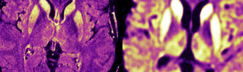
Creutzfeldt-Jakob Disease
Dementia
Creutzfeldt-Jakob Disease Creutzfeldt-Jakob disease (CJD) is the most common human form of a group of rare, fatal brain disorders known as prion diseases. About Symptoms Diagnosis Causes and risks Treatment About Creutzfeldt-Jakob disease Prion diseases, such as Creutzfeldt-Jakob disease, occur when prion protein, which is found throughout the body but whose normal function isn't yet known, begins folding into an abnormal three-dimensional shape. This shape change gradually triggers prion protein in the brain to fold into the same abnormal shape. Creutzfeldt-Jakob disease causes a type of dementia that gets worse unusually fast. More common causes of dementia, such as Alzheimer's , dementia with Lewy bodies and frontotemporal dementia , typically progress more slowly. Through a process scientists don't yet understand, misfolded prion protein destroys brain cells. Resulting damage leads to rapid decline in thinking and reasoning as well as involuntary muscle movements, confusion, difficulty walking and mood changes. Creutzfeldt-Jakob disease is rare, occurring in about one in 1 million people annually worldwide. Experts generally recognize the following main types of Creutzfeldt-Jakob disease: Sporadic Creutzfeldt-Jakob disease develops spontaneously for no known reason. It accounts for 85% of cases. On average, sporadic Creutzfeldt-Jakob disease first appears between ages 60 and 65. Familial Creutzfeldt-Jakob disease is caused by certain changes in the chromosome 20 gene coding the biological blueprint for prion protein. People who develop familial Creutzfeldt-Jakob disease do so because they inherited the genetic changes from a parent. Familial Creutzfeldt-Jakob disease accounts for about 10-15% percent of cases. It develops, on average, at a younger age than sporadic Creutzfeldt-Jakob disease, with some genetic types appearing as early as ages 20 to 40. Acquired Creutzfeldt-Jakob disease results from exposure to an external source of abnormal prion protein. These sources are estimated to account for about 1% of Creutzfeldt-Jakob disease cases. The two most common outside sources are: Medical procedures involving instruments used in neurosurgery, growth hormone from human sources or certain transplanted human tissues, including corneas (the clear outer covering of the eye) and dura mater (the fibrous membrane covering the brain and spinal cord). This type of acquired CJD is also known as iatrogenic CJD (iCJD). The risk of iCJD from medical procedures has been greatly reduced by improved neurosurgical instruments sterilization techniques, new single-use instruments and synthetic sources of growth hormone and dura mater. Meat or other products from cattle infected with bovine spongiform encephalopathy (BVE) or "mad cow disease," recognized in the mid-1990s as the cause of variant CJD (vCJD). Scientists traced this new type of CJD to consumption of beef from cattle whose feed included processed brain tissue from other animals. Since then, experts have diagnosed about 200 cases of vCJD, primarily in the United Kingdom and other European countries. Variant CJD tends to occur at a younger age than sporadic or familial forms, sometimes even in teenagers. New cases of vCJD have slowed significantly, most likely due to changes in animal feeding practices. Chronic wasting disease This prion disease is similar to mad cow disease that's been found in wild deer, elk and moose. According to the Centers for Disease Control (CDC) , there's no evidence to date that the disease has been transmitted to humans. Symptoms Specific Creutzfeldt-Jakob disease symptoms experienced by an individual and the order in which they appear can differ significantly. Some common symptoms include: Depression. Agitation, apathy and mood swings. Rapidly worsening confusion. Disorientation. Problems with memory, thinking, planning and judgment. Difficulty walking. Muscle stiffness, twitches and involuntary jerky movements. Vision problems, such as double vision and hallucinations Diagnosis Rapid symptom progression is one of the most important clues that a person may have Creutzfeldt-Jakob disease. There is no single test — or any combination of tests — that can conclusively diagnose sporadic Creutzfeldt-Jakob disease in a living person, but the following tests may help determine whether an individual has Creutzfeldt-Jakob disease: Electroencephalogram (EEG) measures the brain's patterns of electrical activity similar to the way an electrocardiogram (ECG) measures the heart's electrical activity. Brain magnetic resonance imaging (MRI) can detect certain brain changes consistent with Creutzfeldt-Jakob disease. Lumbar puncture (spinal tap) tests spinal fluid for the presence of certain proteins. Protein misfolding cyclic amplification (PMCA): PMCA is an amplification technique for the detection of misfolded protein aggregates. Causes and risks Sporadic CJD has no known cause. Most scientists believe the disease begins when prion protein somewhere in the brain spontaneously misfolds, triggering a "domino effect" that misfolds prion protein throughout the brain. Genetic variation in the prion protein gene at a location called "codon 129" may increase risk of this spontaneous misfolding. Variation at codon 129 in the prion protein gene may also play a role in making people susceptible to acquired CJD from external sources. Scientists don’t yet know why acquired CJD seems to be transmitted through such a limited number of external sources. Researchers have found no evidence that the abnormal protein is commonly transmitted through sexual activity or blood transfusions, although a few cases of vCJD seem to have been spread through blood transfusions. Professionals who regularly encounter blood from a human or animal, such as surgeons, pathologists or butchers, have not been shown to have a higher-than-normal risk through occupational exposure. Familial CJD is caused by variations in the prion protein gene that increase the likelihood an individual will develop CJD. Researchers have identified more than 50 prion protein mutations in those with inherited CJD. Genetic testing can determine whether family members at risk have inherited a CJD-causing mutation. Experts strongly recommend professional genetic counseling both before and after genetic testing for hereditary CJD. Age has an influence on sporadic CJD, which tends to develop later in life, usually around age 60. Onset of familial CJD occurs slightly earlier and vCJD has affected people at a much younger age, usually in their late 20s. Chronic wasting disease is a prion disease similar to mad cow disease that’s been found in wild deer, elk and moose in certain U.S. states, Canadian provinces, Korea and Norway. According to the U.S. Centers for Disease Control and Prevention (CDC), there’s no evidence to date that chronic wasting disease has been transmitted to humans, including hunters who eat meat from affected animals. There’s also no evidence that rates of CJD have increased in states or provinces where chronic wasting disease has been identified. Additional studies are under way to understand what risk, if any, chronic wasting disease poses to humans. The CDC recommends that hunters who plan to eat meat from deer, elk or moose in areas where chronic wasting disease occurs consider having the meat tested by their local state wildlife agency. The CDC also recommends wearing gloves while field dressing these animals to avoid handling the brain or spinal column. Treatment and outcomes There is no treatment that can slow or stop the underlying brain cell destruction caused by Creutzfeldt-Jakob disease and other prion diseases. Various drugs have been tested but have not shown any benefit. Clinical studies of potential Creutzfeldt-Jakob disease treatments are complicated by the rarity of the disease and its rapid progression. Current therapies focus on treating symptoms and on supporting individuals and families coping with Creutzfeldt-Jakob disease. Doctors may prescribe painkillers such as opiates to treat pain if it occurs. Muscle stiffness and twitching may be treated with muscle-relaxing medications or antiseizure drugs. In the later stages of the disease, individuals with Creutzfeldt-Jakob disease become completely dependent on others for their daily needs and comfort. Creutzfeldt-Jakob disease progresses rapidly. Those affected lose their ability to move or speak and require full-time care to meet their daily needs. An estimated 90 percent of those diagnosed with sporadic Creutzfeldt-Jakob disease die within one year. Those affected by familial Creutzfeldt-Jakob disease tend to develop the disorder at an earlier age and survive somewhat longer than those with the sporadic form, as do those diagnosed with variant Creutzfeldt-Jakob disease. Scientists have not yet learned the reason for these differences in survival. Help is available Creutzfeldt-Jakob Disease Foundation is a nonprofit organization that offers support, information and guidance to those dealing with Creutzfeldt-Jakob disease. Call the Foundation at 800.659.1991. The Alzheimer's Association can help you learn more about Alzheimer's and other dementias, and help you find local support services. Call our 24/7 Helpline at 800.272.3900. Social Security Administration (SSA) has a "compassionate allowance" program in which workers diagnosed with Creutzfeldt-Jakob disease can qualify for Social Security disability benefits. Call the SSA at 800.772.1213. Medline Plus: Creutzfeldt-Jakob disease is a consumer health information service of the U.S. National Library of Medicine and National Institutes of Health (NIH). This gateway page links to resources from NIH agencies, major medical centers and other sources. Source: https://www.alz.org/alzheimers-dementia/what-is-dementia/types-of-dementia/creutzfeldt-jakob-disease
Dementia
Neurological Conditions
Dementia Dementia is not a specific disease. It is a descriptive term for a collection of symptoms that can be caused by a number of disorders that affect the brain. People with dementia have significantly impaired intellectual functioning that interferes with normal activities and relationships. They also lose their ability to solve problems and maintain emotional control, and they may experience personality changes and behavioral problems, such as agitation, delusions, and hallucinations. While memory loss is a common symptom of dementia, memory loss by itself does not mean that a person has dementia. Doctors diagnose dementia only if two or more brain functions - such as memory and language skills -- are significantly impaired without loss of consciousness. Some of the diseases that can cause symptoms of dementia are Alzheimer’s disease (AD), vascular dementia, Lewy body dementia, frontotemporal dementia, Huntington’s disease, and Creutzfeldt-Jakob disease. Doctors have identified other conditions that can cause dementia or dementia-like symptoms including reactions to medications, metabolic problems and endocrine abnormalities, nutritional deficiencies, infections, poisoning, brain tumors, anoxia, or hypoxia (conditions in which the brain’s oxygen supply is either reduced or cut off entirely), and heart and lung problems. Although it is common in very elderly individuals, dementia is not a normal part of the aging process. Moreover, recent studies have found that newer brain scans may point to other causes of Dementia in approximately one-third of presumed AD cases, thereby helping avoid an Alzheimer’s disease misdiagnosis, which may lead to better treatment and care.** A fundamental concept to grasp is that the symptoms of Dementia often go beyond memory loss. They can include significant shifts in mood, more falls, disturbed gait (how we walk), and more. In addition, hallucinations, delusions, and paranoia are not uncommon. Click below on the various terms to learn more about both common and more rare conditions, syndromes and diseases, that can cause, or include symptoms leading to Dementia: Dementia-Like Conditions (that may be reversible); Mild Cognitive Impairment (MCI); Alzheimer's Disease (AD or ALZ) Dementia; Mixed Dementia; Vascular Dementia; Young Onset Dementia; Lewy Body Dementia (LBD); Frontotemporal Dementia (FTD); AIDS Dementia Complex (ADC); Huntington's Disease with Dementia; Multiple Sclerosis (MS) with Dementia; Parkinson's Disease (PD) with Dementia; Chronic Traumatic Brain Injury (CTE) Dementia; Traumatic Brain Injury (TBI) with Dementia; Down Syndrome with Dementia; Posterior Cortical Atrophy (PCA); Primary Progressive Aphasia (PPA); Wernicke-Korsakoff Syndrome (WKS) Dementia; Limbic-predominant Age-related TDP-43 Encephalopathy (LATE);*** Creutzfeldt-Jakob Disease (CJD) Dementia; Corticobasal Degeneration (CBD); Progressive Supranuclear Palsy (PSP); CADASIL;*** Sanfilippo Syndrome*** Batten Disease (Childhood Dementia);*** Binswanger Disease.*** Cerebral Amyloid Angiopathy (CAA)*** Various Childhood Dementias*** Adult-Onset Leukoencephalopathy*** See All Definitions: https://www.dementiasociety.org/definitions Additional Resources: The links below will open websites in a separate web browser page so that you can keep your place here. Antidote- Dementia Clinical Drug Trial Selector - Be taken through a series of questions to find the trial that fits. CenterWatch- Dementia Clinical Drug Trials Database - Search to find trials in your area, and sign up for email alerts. National Institutes of Health- Alzheimer's - Detailed epidemiology (incidence, distribution, and possible control) of Alzheimer's Disease Dementia. U.S. Department of Health and Human Services- Institute on Aging - General information on U.S. government research on aging and related health issues. U.S. Department of Health and Human Services- Brain Health Info - The Association for Community Living (ACL) provides a resource on brain health. World Health Organization- Dementias Fact Sheet - A global Dementia fact sheet produced by a branch of the United Nations. Dementia Prevention, Intervention, and Care- Commission - The Lancet is an independent, international weekly general medical journal founded in 1823. The journal strives to make science widely available so that medicine can serve and transform society and positively impact the lives of people. Brain Bank Donation- Locator - If you wish to help others through Dementia research, consider giving the gift of your brain to science when you die. CDC- Dementia Care Community Stati tics - The U.S. Department of Health and Human Services' Centers for Disease Control (CDC) has prepared a statistical overview (2010) of care communities across America. CMS- Center for Medicare & Medicaid Services - If you have a complaint to make about care for someone living with Dementia who is a resident of a state or federally-certified healthcare facility, you can find the appropriate state contact phone number and web address here. See Care Education: https://www.dementiasociety.org/education Don't see what you're looking for? - Please contact us. Additional Information Donate Home Bequests About Information Resources Memorials Search Source: https://www.dementiasociety.org/
Dementia
Dementia
Dementia Dementia is not a specific disease. It is a descriptive term for a collection of symptoms that can be caused by a number of disorders that affect the brain. People with dementia have significantly impaired intellectual functioning that interferes with normal activities and relationships. They also lose their ability to solve problems and maintain emotional control, and they may experience personality changes and behavioral problems, such as agitation, delusions, and hallucinations. While memory loss is a common symptom of dementia, memory loss by itself does not mean that a person has dementia. Doctors diagnose dementia only if two or more brain functions - such as memory and language skills -- are significantly impaired without loss of consciousness. Some of the diseases that can cause symptoms of dementia are Alzheimer’s disease (AD), vascular dementia, Lewy body dementia, frontotemporal dementia, Huntington’s disease, and Creutzfeldt-Jakob disease. Doctors have identified other conditions that can cause dementia or dementia-like symptoms including reactions to medications, metabolic problems and endocrine abnormalities, nutritional deficiencies, infections, poisoning, brain tumors, anoxia, or hypoxia (conditions in which the brain’s oxygen supply is either reduced or cut off entirely), and heart and lung problems. Although it is common in very elderly individuals, dementia is not a normal part of the aging process. Moreover, recent studies have found that newer brain scans may point to other causes of Dementia in approximately one-third of presumed AD cases, thereby helping avoid an Alzheimer’s disease misdiagnosis, which may lead to better treatment and care.** A fundamental concept to grasp is that the symptoms of Dementia often go beyond memory loss. They can include significant shifts in mood, more falls, disturbed gait (how we walk), and more. In addition, hallucinations, delusions, and paranoia are not uncommon. Click below on the various terms to learn more about both common and more rare conditions, syndromes and diseases, that can cause, or include symptoms leading to Dementia: Dementia-Like Conditions (that may be reversible); Mild Cognitive Impairment (MCI); Alzheimer's Disease (AD or ALZ) Dementia; Mixed Dementia; Vascular Dementia; Young Onset Dementia; Lewy Body Dementia (LBD); Frontotemporal Dementia (FTD); AIDS Dementia Complex (ADC); Huntington's Disease with Dementia; Multiple Sclerosis (MS) with Dementia; Parkinson's Disease (PD) with Dementia; Chronic Traumatic Brain Injury (CTE) Dementia; Traumatic Brain Injury (TBI) with Dementia; Down Syndrome with Dementia; Posterior Cortical Atrophy (PCA); Primary Progressive Aphasia (PPA); Wernicke-Korsakoff Syndrome (WKS) Dementia; Limbic-predominant Age-related TDP-43 Encephalopathy (LATE);*** Creutzfeldt-Jakob Disease (CJD) Dementia; Corticobasal Degeneration (CBD); Progressive Supranuclear Palsy (PSP); CADASIL;*** Sanfilippo Syndrome*** Batten Disease (Childhood Dementia);*** Binswanger Disease.*** Cerebral Amyloid Angiopathy (CAA)*** Various Childhood Dementias*** Adult-Onset Leukoencephalopathy*** See All Definitions: https://www.dementiasociety.org/definitions Additional Resources: The links below will open websites in a separate web browser page so that you can keep your place here. Antidote- Dementia Clinical Drug Trial Selector - Be taken through a series of questions to find the trial that fits. CenterWatch- Dementia Clinical Drug Trials Database - Search to find trials in your area, and sign up for email alerts. National Institutes of Health- Alzheimer's - Detailed epidemiology (incidence, distribution, and possible control) of Alzheimer's Disease Dementia. U.S. Department of Health and Human Services- Institute on Aging - General information on U.S. government research on aging and related health issues. U.S. Department of Health and Human Services- Brain Health Info - The Association for Community Living (ACL) provides a resource on brain health. World Health Organization- Dementias Fact Sheet - A global Dementia fact sheet produced by a branch of the United Nations. Dementia Prevention, Intervention, and Care- Commission - The Lancet is an independent, international weekly general medical journal founded in 1823. The journal strives to make science widely available so that medicine can serve and transform society and positively impact the lives of people. Brain Bank Donation- Locator - If you wish to help others through Dementia research, consider giving the gift of your brain to science when you die. CDC- Dementia Care Community Stati tics - The U.S. Department of Health and Human Services' Centers for Disease Control (CDC) has prepared a statistical overview (2010) of care communities across America. CMS- Center for Medicare & Medicaid Services - If you have a complaint to make about care for someone living with Dementia who is a resident of a state or federally-certified healthcare facility, you can find the appropriate state contact phone number and web address here. See Care Education: https://www.dementiasociety.org/education Don't see what you're looking for? - Please contact us. Additional Information Donate Home Bequests About Information Resources Memorials Search Source: https://www.dementiasociety.org/
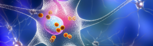
Dementia with Lewy Bodies
Dementia
Dementia with Lewy Bodies Dementia with Lewy bodies (DLB) is a type of progressive dementia that leads to a decline in thinking, reasoning and independent function. Its features may include spontaneous changes in attention and alertness, recurrent visual hallucinations, REM sleep behavior disorder, and slow movement, tremors or rigidity. About Symptoms Diagnosis Causes and risks Treatment About dementia with Lewy bodies Dementia with Lewy bodies is one of the causes of dementia , alongside other types of dementia like Alzheimer's disease and vascular dementia . "Lewy body dementia" (LBD) is a broad term that includes both dementia with Lewy bodies and Parkinson’s disease dementia. Here, we focus on the specific brain disorder called "dementia with Lewy bodies" (DLB). The hallmark brain abnormalities linked to DLB are named after Frederich H. Lewy, M.D., the neurologist who discovered them while working in Dr. Alois Alzheimer's laboratory during the early 1900s. Alpha-synuclein protein, the chief component of Lewy bodies, is found widely in the brain, but its normal function isn't yet known. Lewy bodies may also be found in other types of dementia, including Alzheimer's disease dementia , and are a primary brain abnormality in Parkinson's disease dementia . Many people with Parkinson's eventually develop problems with thinking and reasoning, and many people with DLB experience movement symptoms like hunched posture, rigid muscles, a shuffling walk and trouble initiating movement. This overlap in symptoms and other evidence suggest that dementia with Lewy bodies, Parkinson's disease, and Parkinson's disease dementia may be linked to the same underlying abnormalities in how the brain processes the protein alpha-synuclein. Many people with both DLB and Parkinson's dementia also have plaques and tangles — hallmark brain changes linked to Alzheimer's disease. When people have brain changes of more than one type of dementia, they are said to have mixed dementia. Learn more: Mixed Dementia . Symptoms of dementia with Lewy bodies Core symptoms of dementia with Lewy bodies include: Changes in thinking and reasoning. Fluctuating cognition that is delirium-like. Recurrent well-formed visual hallucinations. REM sleep behavior disorder that involves acting out dreams. Spontaneous parkinsonism with slowness of movement, rest tremor, or rigidity. Other symptoms may include: Trouble interpreting visual information. Malfunctions of the "automatic" (autonomic) nervous system, which controls automatic functions of the body, such as sweating, blood pressure, heart rate, digestion and sexual response. Memory loss that may be significant, but less prominent than in Alzheimer's. Learn more about these and other possible symptoms from the Lewy Body Dementia Association . Families facing dementia need you Donate today so we can continue advancing critical research and providing free 24/7 support to those who depend on it. Donate Now Diagnosis of dementia with Lewy bodies As with other types of dementia , there is no single test that can conclusively diagnose dementia with Lewy bodies. Today, DLB is a "clinical" diagnosis, which means it represents a doctor's best professional judgment about the reason for a person's symptoms. The only way to conclusively diagnose DLB is through a postmortem autopsy. Most experts believe that dementia with Lewy bodies and Parkinson's disease dementia are two different expressions of the same underlying problems with brain processing of the protein alpha-synuclein. They recommend continuing to diagnose DLB and Parkinson's disease dementia as separate disorders. The diagnosis is DLB when a person experiences dementia either before, at the same time as, or within one year of the onset of symptoms of Parkinson’s disease. In some cases of DLB, symptoms of Parkinson’s disease, like changes in movement, may never occur. The diagnosis is Parkinson's disease dementia when a person experiences dementia at least one year (and usually several years) after the onset of symptoms of Parkinson’s disease. Parkinson's disease symptoms may include changes in movement like a tremor. Since Lewy bodies usually coexist with Alzheimer's brain changes , it may sometimes be hard to distinguish DLB from Alzheimer's disease, especially in the early stages. Key differences between Alzheimer's and dementia with Lewy bodies Memory loss tends to be a more prominent symptom in early Alzheimer's than in early DLB. However, advanced DLB may cause memory problems in addition to its more typical effects on judgment, planning and visual perception. Movement symptoms are more likely to be an important cause of disability early in DLB than in Alzheimer's. However, Alzheimer's can cause problems with walking, balance and getting around as it progresses to moderate and severe stages. Hallucinations and misidentification of familiar people are significantly more frequent in early-stage DLB than in Alzheimer's. REM sleep disorder is more common in early DLB than in Alzheimer's. Disruption of the autonomic nervous system — such as causing a blood pressure drop on standing, dizziness, falls and urinary incontinence — is much more common in early DLB than in Alzheimer's. Causes and risks Researchers have not yet identified any specific causes of dementia with Lewy bodies. Most people diagnosed with DLB have no family history of the disorder, and no genes linked to DLB have been conclusively identified. Treatment and outcomes There are no treatments that can slow or stop the brain cell damage caused by dementia with Lewy bodies. Current strategies focus on helping symptoms. If your treatment plan includes medications, it's important to work closely with your physician to identify the drugs that work best for you and the most effective doses. Treatment considerations involving medications include the following issues: Cholinesterase inhibitor drugs are a common approach for addressing thinking changes in Alzheimer's. They also may help certain DLB symptoms. Antipsychotic drugs should be used with extreme caution in Lewy body dementia, including both dementia with Lewy bodies and Parkinson's disease dementia. Although physicians sometimes prescribe these drugs for behavioral symptoms that can occur in Alzheimer's , they may cause serious side effects in as many as 50% of those with Lewy body dementia . Side effects may include sudden changes in consciousness, impaired swallowing, acute confusion, episodes of delusions or hallucinations, or appearance or worsening of Parkinson's symptoms. Antidepressants may be used to treat depression , which is common with DLB, Parkinson's disease dementia and Alzheimer's. The most commonly used antidepressants are selective serotonin reuptake inhibitors (SSRIs). Like other types of dementia that destroy brain cells , dementia with Lewy bodies gets worse over time and shortens lifespan. Help is available Lewy Body Dementia Association (LBDA) is a nonprofit organization providing information and assistance to individuals with the disease, caregivers and medical professionals. Call LBDA at 800.539.9767. The Alzheimer's Association can help you learn more about Alzheimer's and other dementias, and help you find local support services. Call our 24/7 Helpline at 800.272.3900. The Social Security Administration (SSA) has a "compassionate allowance" program in which workers diagnosed with Lewy body dementia can qualify for Social Security disability benefits. Call the SSA at 800.772.1213. The National Institute on Aging provides details on the signs and symptoms of Lewy body dementia, as well as treatment and care options. Source: https://www.alz.org/alzheimers-dementia/what-is-dementia/types-of-dementia/dementia-with-lewy-bodies

Demyelinating Syndromes (Optic Neuritis, Transverse Myelitis, ADEM)
Neurological Conditions
Demyelinating Syndromes (Optic Neuritis, Transverse Myelitis, ADEM) & Neuromyelitis Optica Spectrum Disorders (NMOSD) Demyelinating syndromes represent a group of inflammatory autoimmune attacks against the central nervous system (CNS), leading to acute or subacute neurological symptoms involving vision, movement, sensation, coordination, and bladder/bowel function. These episodes may occur as isolated events or as part of a broader neuroimmunological disease such as multiple sclerosis (MS) or neuromyelitis optica spectrum disorder (NMOSD). The most common isolated demyelinating syndromes include: Optic Neuritis (ON) – inflammation of the optic nerve causing vision loss and pain with eye movement. Transverse Myelitis (TM) – inflammation across one segment of the spinal cord causing weakness, numbness, or bladder dysfunction. Acute Disseminated Encephalomyelitis (ADEM) – widespread inflammation of brain and spinal cord, usually following infection and more common in children. These conditions must also be distinguished from Neuromyelitis Optica Spectrum Disorders (NMOSD) —a severe antibody-mediated disease primarily affecting the optic nerves and spinal cord, associated with AQP4-IgG or MOG-IgG antibodies. Prompt recognition and treatment are essential because early immunotherapy can dramatically improve long-term outcomes and prevent irreversible neurological damage. Questions to Ask Your Doctor • Is this episode isolated, or does it suggest MS, NMOSD, or MOG-associated disease? • What imaging findings or antibodies support the diagnosis? • Do I need a spinal tap, MRI of brain/spine, or visual evoked potentials? • Should we test for AQP4-IgG and MOG-IgG antibodies? • What is the risk of relapse, and how do we prevent new attacks? • Do I need high-dose steroids, plasma exchange, or long-term immunotherapy? • How do we monitor recovery of vision, strength, or sensation? • What symptoms require urgent reevaluation? • Can rehabilitation improve mobility, balance, or daily functioning? Overview Demyelinating syndromes arise when the immune system attacks the myelin coating of CNS nerves. Depending on the location of inflammation, individuals may experience vision loss, limb weakness, sensory deficits, gait disturbance, bowel/bladder problems, or encephalopathy. These episodes fall into two broad categories: Monophasic demyelination — single event, may fully recover (e.g., typical optic neuritis, some cases of TM, ADEM). Relapsing or evolving disease — part of a chronic autoimmune condition such as MS, NMOSD, or MOGAD. Understanding the underlying pathology is essential because prognosis and treatment strategies differ substantially. DEMYELINATING SYNDROMES 1. Optic Neuritis (ON) Clinical Features • Subacute, often unilateral vision loss • Pain with eye movement • Loss of color vision (dyschromatopsia) • Central scotoma or blurred vision • Afferent pupillary defect What Makes ON More Likely? • Young adult onset • Vision worsens over 1–7 days • Pain with eye movement • MRI showing optic nerve enhancement Causes • MS-associated ON (most common) • MOG antibody–associated disease (MOGAD) • NMOSD (AQP4-IgG–positive) • Post-infectious or idiopathic 2. Transverse Myelitis (TM) Clinical Features • Bilateral leg or arm weakness • Numbness, tingling, or sensory level on torso • Bowel or bladder dysfunction • Back pain • Gait disturbance Patterns • Partial TM — often MS • Longitudinally extensive TM (LETM, >3 vertebral segments) — highly suggestive of NMOSD or MOGAD 3. Acute Disseminated Encephalomyelitis (ADEM) Clinical Features • Encephalopathy (confusion, lethargy) • Multifocal neurological deficits • Ataxia, weakness, seizures • Often follows viral illness or vaccination • More common in children MRI Pattern • Large, diffuse, poorly defined lesions in brain and sometimes spinal cord • Often bilateral and symmetric NEUROMYELITIS OPTICA SPECTRUM DISORDERS (NMOSD) NMOSD is a severe autoimmune disorder in which antibodies target aquaporin-4 (AQP4) channels on astrocytes, leading to intense inflammation of optic nerves and spinal cord. Without rapid treatment, NMOSD attacks can cause profound, irreversible disability. Key Features Suggesting NMOSD • Recurrent optic neuritis • Bilateral or severe ON • Transverse myelitis involving ≥3 vertebral segments • Intractable hiccups, nausea (area postrema syndrome) • Persistent, severe pain • Poor recovery from initial attack Types AQP4-IgG–positive NMOSD — classic form. Seronegative NMOSD — diagnosis based on clinical features + MRI (rare). MOG-IgG Disease (MOGAD) — distinct entity that can mimic NMOSD or MS but has better recovery and different treatment. Signs and Symptoms Optic Neuritis • Blurred or dim vision • Pain with eye movement • Color desaturation • Visual field loss Transverse Myelitis • Limb weakness • Sensory changes • Heightened reflexes or spasticity • Bladder urgency or retention ADEM • Altered mental status • Seizures • Multifocal deficits • Headache or fever NMOSD • Severe, recurrent ON • LETM with marked weakness • Persistent vomiting/hiccups (area postrema) • Severe neuropathic pain • Poor spontaneous recovery Exams and Tests 1. MRI Brain and Spine • Optic nerve enhancement (ON) • Cervical or thoracic lesions (TM) • LETM (>3 segments) → NMOSD/MOGAD • Large ADEM lesions 2. Lumbar Puncture • Oligoclonal bands → suggests MS • Markedly elevated protein in TM • Absence of OCBs with LETM → raises concern for NMOSD 3. Serum Antibody Testing (Mandatory) • AQP4-IgG — NMOSD • MOG-IgG — MOG-associated demyelination 4. Evoked Potentials • Visual evoked potentials for ON • Somatosensory evoked potentials for myelitis 5. Routine Labs To exclude mimics: • B12, copper • ANA, ENA • Thyroid function • Lyme, syphilis, HIV • ESR/CRP 6. Advanced Biomarkers • Neurofilament light chain (NfL) — axonal injury • GFAP — astrocytic injury (elevated in NMOSD) Treatment Treatment depends on whether the event is isolated or part of chronic disease. Acute Attack Management (All Forms) • High-dose IV methylprednisolone (first-line) • Plasma exchange (PLEX) for severe attacks or steroid non-responders • IVIG (select cases, especially MOGAD or pediatric demyelination) Early treatment can significantly improve vision, mobility, and long-term function. Long-Term Disease Prevention For MS-like monophasic ON or TM Treatment varies depending on recurrence risk and MRI features. For NMOSD (AQP4-IgG positive) — Always requires long-term therapy • Eculizumab (C5 inhibitor) • Inebilizumab (anti-CD19) • Satralizumab (IL-6 receptor blocker) These therapies dramatically reduce relapse risk and disability progression. For MOGAD • Long-term therapy only if relapsing (steroids, IVIG, mycophenolate, rituximab) • Generally better recovery than NMOSD Symptom Management • Neuropathic pain medications • Bowel/bladder management • Spasticity treatment (baclofen, tizanidine) • Mobility aids and PT/OT • Vision rehabilitation for ON Living With Demyelinating Syndromes & NMOSD Individuals often improve significantly with early treatment, structured follow-up, and multidisciplinary support. Effective strategies: • Regular MRI monitoring • Early recognition of relapse symptoms • Avoiding overheating and infections • Vitamin D optimization • Anti-inflammatory nutrition • Stress reduction and sleep optimization NMOSD requires lifelong immunotherapy to prevent relapses, while many cases of ON, TM, or ADEM are monophasic. Did You Know? • Transverse myelitis that spans >3 vertebral segments is highly suggestive of NMOSD. • MOGAD often affects children and young adults and has excellent recovery with steroids. • AQP4 antibodies target astrocytes—not myelin—leading to secondary demyelination. • ADEM is typically a one-time event and often follows infection. • Optic neuritis associated with MS usually recovers well within weeks to months. References Wingerchuk DM, Banwell B, Bennett JL, et al. International Consensus Diagnostic Criteria for Neuromyelitis Optica Spectrum Disorders. Neurology. 2015;85(2):177-189. [1] https://pubmed.ncbi.nlm.nih.gov/26092914 International consensus criteria for NMOSD: six core clinical characteristics (optic neuritis, acute myelitis, area postrema syndrome, brainstem syndrome, narcolepsy, cerebral syndrome); AQP4-IgG testing required; diagnostic algorithm Papadopoulos MC, Verkman AS. Aquaporin 4 and Neuromyelitis Optica. Lancet Neurol. 2012;11(6):535-544. https://pubmed.ncbi.nlm.nih.gov/22608667 AQP4 water channels on astrocyte foot processes; AQP4-IgG binding triggers complement activation, granulocyte infiltration, astrocyte destruction, secondary demyelination; longitudinally extensive spinal cord lesions characteristic Weinshenker BG, Wingerchuk DM. Neuromyelitis Optica: Clinical Syndrome and the NMO-IgG Autoantibody Marker. Curr Top Microbiol Immunol. 2008;318:343-356. https://pubmed.ncbi.nlm.nih.gov/18219825 Clinical features distinguishing NMOSD from MS: severe attacks with poor recovery, bilateral/recurrent optic neuritis, longitudinally extensive transverse myelitis ≥3 vertebral segments, area postrema syndrome Levy M, Fujihara K, Palace J. New Therapies for Neuromyelitis Optica Spectrum Disorder. Lancet Neurol. 2021;20(1):60-67. [2] https://pubmed.ncbi.nlm.nih.gov/33340490 Review of NMOSD therapies: eculizumab (complement C5 inhibitor), inebilizumab (anti-CD19 B-cell depletion), satralizumab (IL-6 receptor blockade); all reduce annualized relapse rate by 73-79% Flanagan EP, Cabre P, Weinshenker BG, et al. Epidemiology of Aquaporin-4 Autoimmunity and Neuromyelitis Optica Spectrum. Ann Neurol. 2016;79(5):775-783. https://pubmed.ncbi.nlm.nih.gov/26990317 NMOSD epidemiology: prevalence 0.5-10 per 100,000; female:male ratio 9:1; higher prevalence in non-white populations; median age onset 39 years; 10% seronegative NMOSD Treatment Trials (3 references): Pittock SJ, Berthele A, Fujihara K, et al. Eculizumab in Aquaporin-4-Positive Neuromyelitis Optica Spectrum Disorder (N-MOmentum): A Double-Blind, Randomised Placebo-Controlled Phase 3 Trial. Lancet. 2019;394(10206):1352-1363. [1] https://pubmed.ncbi.nlm.nih.gov/31495497 N-MOmentum trial: eculizumab reduced adjudicated relapse risk by 94% vs placebo in AQP4-IgG+ NMOSD; complement C5 inhibitor blocks terminal complement activation Cree BAC, Bennett JL, Kim HJ, et al. Inebilizumab for the Treatment of Neuromyelitis Optica Spectrum Disorder (N-MOmentum): A Double-Blind, Randomised Placebo-Controlled Phase 2/3 Trial. Lancet. 2019;394(10206):1352-1363. [2] https://pubmed.ncbi.nlm.nih.gov/31495497 N-MOmentum trial: inebilizumab (anti-CD19) reduced attack risk by 73% in AQP4-IgG+ patients, 78% in all NMOSD; B-cell depletion mechanism Yamamura T, Kleiter I, Fujihara K, et al. Trial of Satralizumab in Neuromyelitis Optica Spectrum Disorder. N Engl J Med. 2019;381(22):2114-2124. https://www.nejm.org/doi/full/10.1056/NEJMoa1901747 SAkuraSky trial: satralizumab (IL-6 receptor blocker) reduced relapse risk by 62% overall, 79% in AQP4-IgG+ patients; subcutaneous administration; can be used as monotherapy MOG Antibody-Associated Disease (4 references): Jarius S, Paul F, Aktas O, et al. MOG Encephalomyelitis: International Recommendations on Diagnosis and Antibody Testing. J Neuroinflammation. 2018;15(1):134. https://pubmed.ncbi.nlm.nih.gov/29724224 MOG-IgG diagnostic recommendations: cell-based assays required (not ELISA); testing indicated for optic neuritis, transverse myelitis, ADEM, NMOSD-like presentations; repeat testing if initially negative Banwell B, Bennett JL, Marignier R, et al. Diagnosis of Myelin Oligodendrocyte Glycoprotein Antibody-Associated Disease: International MOGAD Panel Proposed Criteria. Lancet Neurol. 2023;22(3):268-282. https://pubmed.ncbi.nlm.nih.gov/36657752 - 2023 international diagnostic criteria for MOGAD: clinical attack + MOG-IgG (cell-based assay) + reasonable exclusion of alternatives; distinct from MS and NMOSD; 25-50% monophasic 11. Cobo-Calvo A, Ruiz A, Maillart E, et al. Clinical Spectrum and Prognostic Value of CNS MOG Autoimmunity in Adults: The MOGADOR Study. Neurology. 2018;90(21):e1858-e1869. https://pubmed.ncbi.nlm.nih.gov/29695598 - MOGADOR study: adult MOG-IgG disease phenotypes include optic neuritis (58%), myelitis (18%), ADEM (6%), brainstem/cerebral cortical encephalitis; 50% relapsing; better recovery than AQP4-NMOSD 12. Sato DK, Callegaro D, Lana-Peixoto MA, et al. Distinction Between MOG Antibody-Positive and AQP4 Antibody-Positive NMO Spectrum Disorders. Neurology. 2014;82(6):474-481. https://pubmed.ncbi.nlm.nih.gov/24415568 - Distinguishing features: MOG-IgG associated with simultaneous bilateral optic neuritis, conus involvement in myelitis, better recovery, less female predominance vs AQP4-NMOSD Optic Neuritis (3 references): Bennett JL, de Seze J, Lana-Peixoto M, et al. Neuromyelitis Optica and Multiple Sclerosis: Seeing Differences Through Optical Coherence Tomography. Mult Scler. 2015;21(6):678-688. https://pubmed.ncbi.nlm.nih.gov/25662345 - OCT in optic neuritis: MS-associated ON shows mild-moderate RNFL thinning; NMOSD shows severe microcystic macular edema and profound RNFL loss; MOG intermediate 14. Toosy AT, Mason DF, Miller DH. Optic Neuritis. Lancet Neurol. 2014;13(1):83-99. https://pubmed.ncbi.nlm.nih.gov/24331794 - Comprehensive ON review: subacute monocular vision loss with pain; high-dose IV methylprednisolone accelerates recovery but doesn't affect final visual outcome; 50% MS risk at 15 years if MRI lesions present 15. Beck RW, Cleary PA, Anderson MM, et al. A Randomized, Controlled Trial of Corticosteroids in the Treatment of Acute Optic Neuritis. N Engl J Med. 1992;326(9):581-588. https://www.nejm.org/doi/full/10.1056/NEJM199202273260901 - Landmark Optic Neuritis Treatment Trial: IV methylprednisolone (1g/day × 3 days) accelerated visual recovery; oral prednisone alone increased recurrence risk; established standard of care Transverse Myelitis & ADEM (3 references): West TW, Hess C, Cree BAC. Acute Transverse Myelitis: Demyelinating, Inflammatory, and Infectious Myelopathies. Semin Neurol. 2012;32(2):97-113. https://pubmed.ncbi.nlm.nih.gov/22961311 - Diagnostic approach to transverse myelitis: distinguish partial vs complete; short vs longitudinally extensive (LETM ≥3 segments suggests NMOSD/MOGAD); parenchymal vs meningeal enhancement patterns 17. Flanagan EP, Weinshenker BG, Krecke KN, et al. Short Myelitis Lesions in Aquaporin-4-IgG-Positive Neuromyelitis Optica Spectrum Disorders. JAMA Neurol. 2015;72(1):81-87. https://pubmed.ncbi.nlm.nih.gov/25384099 - 14% of AQP4-IgG+ NMOSD patients present with short cord lesions (<3 segments); can evolve to LETM; clinical severity and CSF pleocytosis help distinguish from MS 18. Pohl D, Alper G, Van Haren K, et al. Acute Disseminated Encephalomyelitis: Updates on an Inflammatory CNS Syndrome. Neurology. 2016;87(9 Suppl 2):S38-45. https://pubmed.ncbi.nlm.nih.gov/27572860 - ADEM diagnostic criteria: encephalopathy (required) + multifocal CNS symptoms; MRI shows large, poorly demarcated lesions; typically monophasic; often post-infectious; distinguish from first MS attack Treatment & Recovery (2 references): Kleiter I, Gahlen A, Borisow N, et al. Neuromyelitis Optica: Evaluation of 871 Attacks and 1,153 Treatment Courses. Ann Neurol. 2016;79(2):206-216. https://pubmed.ncbi.nlm.nih.gov/26537743 - Large NMOSD treatment study: high-dose corticosteroids first-line (77% attacks); plasma exchange for steroid failures reduced disability; early treatment (within 7 days) associated with better outcomes 20. Bonnan M, Valentino R, Debeugny S, et al. Short Delay to Initiate Plasma Exchange Is the Strongest Predictor of Outcome in Severe Attacks of NMO Spectrum Disorders. J Neurol Neurosurg Psychiatry. 2018;89(4):346-351. https://pubmed.ncbi.nlm.nih.gov/29101261 • Wingerchuk DM et al. International consensus diagnostic criteria for NMOSD. Neurology. 2015; updated reviews 2020–2023. • Flanagan EP. Idiopathic transverse myelitis: emerging biomarkers and phenotypes. Lancet Neurol. 2021. • Sato DK et al. MOG-IgG–associated disease: clinical features and treatment. J Neurol Neurosurg Psychiatry. 2022. • Jarius S, Wildemann B. AQP4-antibody positive NMOSD: current therapies and outcomes. Front Neurol. 2021. • Waldman A et al. ADEM and pediatric demyelinating syndromes. Nat Rev Neurol. 2020. Source: https://www.premierneurohealth.com/

Depression
Neurological Conditions
Depression Depression is a common mental health condition that causes a persistent feeling of sadness and changes in how you think, sleep, eat and act. There are several different types. Depression is treatable — usually with talk therapy, medication or both. Seeking medical help as soon as you have symptoms is essential. What is depression? Depression is a mood disorder that causes a persistent feeling of sadness and loss of interest in things and activities you once enjoyed. It can also cause difficulty with thinking, memory, eating and sleeping . It’s normal to feel sad about or grieve over difficult life situations, such as losing your job or a divorce. But depression is different in that it persists practically every day for at least two weeks and involves other symptoms than sadness alone. There are several types of depressive disorders. Clinical depression, or major depressive disorder, is often just called “depression.” It’s the most severe type of depression. Without treatment, depression can get worse and last longer. In severe cases, it can lead to self-harm or death by suicide . The good news is that treatments can be very effective in improving symptoms. What are the types of depression? The American Psychiatric Association’s Diagnostic Statistical Manual of Mental Disorders, Fifth Edition (DSM-5) classifies depressive disorders as the following: Clinical depression (major depressive disorder) : A diagnosis of major depressive disorder means you’ve felt sad, low or worthless most days for at least two weeks while also having other symptoms such as sleep problems, loss of interest in activities or change in appetite. This is the most severe form of depression and one of the most common forms. Persistent depressive disorder (PDD) : Persistent depressive disorder is mild or moderate depression that lasts for at least two years. The symptoms are less severe than major depressive disorder. Healthcare providers used to call PDD dysthymia. Disruptive mood dysregulation disorder (DMDD) : DMDD causes chronic, intense irritability and frequent anger outbursts in children. Symptoms usually begin by the age of 10. Premenstrual dysphoric disorder (PMDD) : With PMDD, you have premenstrual syndrome (PMS) symptoms along with mood symptoms, such as extreme irritability, anxiety or depression. These symptoms improve within a few days after your period starts, but they can be severe enough to interfere with your life. Depressive disorder due to another medical condition : Many medical conditions can create changes in your body that cause depression. Examples include hypothyroidism , heart disease , Parkinson’s disease and cancer. If you’re able to treat the underlying condition, the depression usually improves as well. There are also specific forms of major depressive disorder, including: Seasonal affective disorder (seasonal depression) : This is a form of major depressive disorder that typically arises during the fall and winter and goes away during the spring and summer. Prenatal depression and postpartum depression : Prenatal depression is depression that happens during pregnancy. Postpartum depression is depression that develops within four weeks of delivering a baby. The DSM refers to these as “major depressive disorder (MDD) with peripartum onset.” Atypical depression : Symptoms of this condition, also known as major depressive disorder with atypical features, vary slightly from “typical” depression. The main difference is a temporary mood improvement in response to positive events (mood reactivity). Other key symptoms include increased appetite and rejection sensitivity. People with bipolar disorder also experience episodes of depression in addition to manic or hypomanic episodes. Who does depression affect? Depression can affect anyone — including children and adults. Women and people assigned female at birth are more likely to have depression than men and people assigned male at birth. Having certain risk factors makes it more likely that you may develop depression. For example, the following conditions are associated with higher rates of depression: Neurodegenerative diseases like Alzheimer’s disease and Parkinson’s disease. Stroke . Multiple sclerosis . Seizure disorders. Cancer. Macular degeneration . Chronic pain . How common is depression? Depression is common. Researchers estimate that nearly 7% of adults in the United States have depression every year. More than 16% of U.S. adults — around 1 in 6 people — will experience depression at some point in their lifetime. However, researchers believe that these estimates are lower than reality, as many people don’t seek medical help for symptoms of depression and don’t receive a diagnosis. Approximately 4.4% of children in the United States have depression. Causes and Risk Factors Clinical depression is a chronic condition, but it usually occurs in episodes, which can last several weeks or months. What are the symptoms of depression? The symptoms of depression can vary slightly depending on the type and can range from mild to severe. In general, symptoms include: Feeling very sad, hopeless or worried. Children and adolescents with depression may be irritable rather than sad. Not enjoying things that used to bring joy. Being easily irritated or frustrated. Eating too much or too little, which may result in weight gain or weight loss. Trouble sleeping ( insomnia ) or sleeping too much ( hypersomnia ). Having low energy or fatigue. Having a difficult time concentrating, making decisions or remembering things. Experiencing physical issues like headache, stomachache or sexual dysfunction . Having thoughts of self-harm or suicide. If you or a loved one are thinking about suicide, dial 988 on your phone to reach the Suicide and Crisis Lifeline. Someone will be available to help you 24/7. What causes depression? Researchers don’t know the exact cause of depression. They think that several factors contribute to its development, including: Brain chemistry : An imbalance of neurotransmitters , including serotonin and dopamine , contributes to the development of depression. Genetics : If you have a first-degree relative (biological parent or sibling) with depression, you’re about three times as likely to develop the condition as the general population. However, you can have depression without a family history of it. Stressful life events : Difficult experiences, such as the death of a loved one, trauma, divorce, isolation and lack of support, can trigger depression. Medical conditions : Chronic pain and chronic conditions like diabetes can lead to depression. Medication : Some medications can cause depression as a side effect. Substance use, including alcohol, can also cause depression or make it worse. Diagnosis and Tests How is depression diagnosed? Healthcare providers diagnose depression based on a thorough understanding of your symptoms, medical history and mental health history. They may diagnose you with a specific type of depression, such as seasonal affective disorder or postpartum depression, based on the context of your symptoms. To receive a diagnosis of depression, you must have five depression symptoms every day, nearly all day, for at least two weeks. Your provider may order medical tests, such as blood tests, to see if any underlying medical conditions are causing your depressive symptoms. Management and Treatment How is depression treated? Depression is one of the most treatable mental health conditions. Approximately 80% to 90% of people with depression who seek treatment eventually respond well to treatment. Treatment options include: Psychotherapy : Psychotherapy (talk therapy) involves talking with a mental health professional. Your therapist helps you identify and change unhealthy emotions, thoughts and behaviors. There are many types of psychotherapy — cognitive behavioral therapy (CBT) is the most common. Sometimes, brief therapy is all you need. Other people continue therapy for several months or years. Medication : Prescription medicine called antidepressants can help change the brain chemistry that causes depression. There are several different types of antidepressants, and it may take time to figure out the one that’s best for you. Some antidepressants have side effects, which often improve with time. If they don’t, talk to your healthcare provider. A different medication may work better for you. Complementary medicine : This involves treatments you may receive along with traditional Western medicine. People with mild depression or ongoing symptoms can improve their well-being with therapies such as acupuncture , massage, hypnosis and biofeedback . Brain stimulation therapy : Brain stimulation therapy can help people who have severe depression or depression with psychosis. Types of brain stimulation therapy include electroconvulsive therapy (ECT) , transcranial magnetic stimulation (TMS) and vagus nerve stimulation (VNS) . There are also things you can do at home to help improve depression symptoms, including: Getting regular exercise. Getting quality sleep (not too little or too much). Eating a healthy diet. Avoiding alcohol, which is a depressant. Spending time with people you care about. Prevention Can I prevent depression? You can’t always prevent depression, but you can help reduce your risk by: Maintaining a healthy sleep routine. Managing stress with healthy coping mechanisms. Practicing regular self-care activities such as exercise, meditation and yoga. If you’ve had depression before, you may be more likely to experience it again. If you have depression symptoms, get help as soon as possible. Outlook/Prognosis What is the prognosis of depression? The prognosis (outlook) of depression varies depending on certain factors, including: Its severity and type. If it’s temporary or long-lasting. If it’s treated or untreated. If you have co-occurring conditions, such as other mood disorders, medical conditions or substance use disorder . With proper diagnosis and treatment, the vast majority of people with depression live healthy, fulfilling lives. Depression can return after you get treatment, though, so it’s important to seek medical help as soon as symptoms begin again. Without treatment, depression can: Become worse. Increase your chance of other health conditions, like dementia . Lead to the worsening of existing health conditions, like diabetes or chronic pain. Lead to self-harm or death. Depression accounts for nearly 40,000 cases of suicide each year in the United States. It’s essential to get medical help as soon as possible if you’re having suicidal thoughts. Call 911 or 988 (the Suicide and Crisis Lifeline) or go to the emergency room. Living with Depression When should I see my healthcare provider about depression? If you have symptoms of depression, see a healthcare provider or mental health professional. They can give you an accurate diagnosis and suggest treatment options. If you’ve started treatment for depression and it isn’t working or you’re having unpleasant side effects, talk to your provider. They can recommend a different treatment plan. A note from Cleveland Clinic Depression is a common condition that affects millions of people every year. Anyone can experience depression — even if there doesn’t seem to be a reason for it. The good news is that depression is treatable. If you have symptoms of depression, talk to your healthcare provider. The sooner you get help, the sooner you can feel better. Source: https://my.clevelandclinic.org/health/diseases/9290-depression
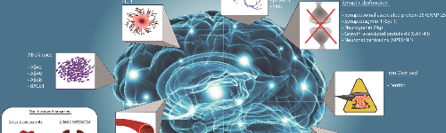
Earlier Diagnosis
Alzheimers
Earlier Diagnosis What if we could diagnose Alzheimer's disease before symptoms started? The hope is, future treatments could then target the disease in its earliest stages, before irreversible brain damage or mental decline has occurred. Research on new strategies for earlier diagnosis is among the most active areas in Alzheimer's science, and funding from the Alzheimer's Association has spurred significant advances and steady progress. Biomarkers for earlier detection Brain imaging/neuroimaging Blood tests Cerebrospinal fluid (CSF) tests Genetic risk profiling Biomarkers for earlier detection Current diagnosis of Alzheimer's disease relies largely on documenting mental decline, at which point, Alzheimer's has already caused severe brain damage. Researchers hope to discover an easy and accurate way to detect Alzheimer's before these devastating symptoms begin. Experts believe that biomarkers (short for "biological markers") offer one of the most promising paths. A biomarker is something that can be measured to accurately and reliably indicate the presence of disease, such as fasting blood glucose (blood sugar) level, which indicates the presence of diabetes if it is 126 mg/dL or higher. Several potential biomarkers are being studied for their ability to indicate early stages of Alzheimer's disease. Examples being studied include beta-amyloid and tau levels in cerebrospinal fluid (CSF) and brain changes detectable by imaging. Recent research suggests that these indicators may change at different stages of the disease process. Before a biomarker can be used in medical clinics, it must be validated, in which multiple studies in large and diverse groups of people establish that it accurately and reliably indicates the presence of disease. Furthermore, the laboratory methods used to measure the biomarker must be shown to be stable and reliable. Currently, there are some FDA-approved tools that, when applicable, can be used to aid in diagnosis of people with symptoms of Alzheimer’s or another dementia (e.g., brain imaging ). Some of these tools have a wealth of research and clinical data to support their use in the clinic (e.g., biomarkers in CSF ), while other emerging biomarkers are promising but still under investigation (e.g., blood tests and genetic risk profiling ). Brain imaging/neuroimaging Neuroimaging is regularly used today for early detection of Alzheimer’s. Research continues to evolve on promising new and advanced brain imaging techniques. Imaging technologies used in Alzheimer's research Structural imaging provides information about the shape, position or volume of brain tissue. Structural techniques include magnetic resonance imaging (MRI) and computed tomography (CT). Functional imaging reveals how well cells in various brain regions are working by showing how actively the cells use sugar or oxygen. Functional techniques include positron emission tomography (PET) and functional MRI (fMRI). Molecular imaging uses highly targeted radiotracers to detect cellular or chemical changes linked to specific diseases. Molecular imaging technologies include PET and fMRI. Structural imaging Having shown that the brains of people with Alzheimer's shrink significantly as the disease progresses, structural imaging research also has shown that shrinkage in specific brain regions such as the hippocampus may be an early sign of Alzheimer's. Scientists now have some agreement on standardized values for brain volume loss (sometimes measured over time) that would indicate the presence of disease or disease progression. Today, a standard workup for Alzheimer's disease may include structural imaging, and these tests are currently used to define pre-existing tissue damage associated with neurodegeneration and to rule out other conditions that may cause symptoms similar to Alzheimer's but require different treatment. Structural imaging such as MRI can reveal tumors, evidence of small or large strokes, damage from severe head trauma, or a buildup of fluid in the brain, as well as detect underlying conditions that may preclude an individual from certain treatments. Functional imaging Functional imaging research suggests that those with Alzheimer's typically have reduced brain cell activity in certain regions. For example, studies with fluorodeoxyglucose (FDG)-positron emission tomography (PET) indicate that Alzheimer's is often associated with reduced use of glucose (sugar) in brain areas important in memory, learning and problem-solving. According to Medicare recommendations, an FDG-PET scan is considered a reasonable test for people with a recent diagnosis of dementia and documented cognitive decline of at least six months who meet diagnostic criteria for both Alzheimer’s and frontotemporal dementia. Molecular imaging Molecular imaging, which also uses PET scans, is among the most active areas of research aimed at finding new approaches to diagnose Alzheimer's in its earliest stages. Molecular strategies may detect biological clues indicating Alzheimer's is under way before the disease changes the brain's structure or function, or takes an irreversible toll on memory, thinking and reasoning. Molecular imaging also may offer a new strategy to monitor disease progression and assess the effectiveness of next-generation, disease-modifying treatments. Several molecular imaging compounds are being studied, and four have been approved for clinical use: Florbetaben (Neuraceq®), Florbetapir (Amyvid®) and Flutemetamol (Vizamyl®) have been approved for detection of beta-amyloid in the brain. Flortaucipir F18 (Tauvid®) has been approved for detection of tau in the brain. Even though amyloid plaques in the brain are a characteristic feature of Alzheimer's disease, their presence alone cannot be used to diagnose the disease. Today, a diagnosis of Alzheimer’s is based on the evaluation of several things, including the presence of amyloid plaques. Your doctor may perform tests to evaluate your memory, order laboratory tests or perform a molecular imaging test (e.g., PET scan) to confirm an Alzheimer’s diagnosis or rule out other diseases that may cause similar symptoms. Cerebrospinal fluid (CSF) tests CSF is a clear fluid that bathes and cushions the brain and spinal cord. Adults have about 1 pint of CSF, which physicians can sample through a minimally invasive procedure called a lumbar puncture, or spinal tap. Research suggests that Alzheimer's disease in early stages may cause changes in CSF levels of multiple markers such as tau and beta-amyloid, two markers that form abnormal brain deposits strongly linked to Alzheimer's. Another potential marker is neurofilament light (NfL), an increased level of which has been found in neurodegenerative diseases such as Alzheimer’s. One challenge researchers face is that analysis of biomarker levels in the same sample can often vary significantly from institution to institution and across different testing platforms. Great strides have been made in standardizing the measurement of these markers in research and clinical care. CSF tests are currently used by dementia specialists to aid in the diagnosis of Alzheimer's, and research continues to develop and standardize new markers that will aid in diagnosis and detection of other dementias. One CSF Amyloid Ratio test, Lumipulse®, received FDA approval and is a new diagnostic tool that clinicians can use to detect amyloid in CSF, which can be predictive of amyloid changes in the brain. The Alzheimer's Association convened a multidisciplinary workgroup and published appropriate use criteria to guide the safe and optimal use of the lumbar puncture procedure and CSF testing for Alzheimer's disease. Blood tests Researchers are investigating whether consistent and measurable changes in blood levels of specific markers may be reliably associated with Alzheimer’s related changes. These markers may include tau, beta-amyloid or other biomarkers the could be measured before and after symptoms appear. An urgent need exists for simple, inexpensive, non-invasive and easily available diagnostic tools such as blood tests to diagnose the disease. These testing technologies would support drug development by helping to identify and follow treatment effectiveness in clinical trial participants and to increase the possibility of early detection, diagnosis and intervention. A blood test would also enable interpretation and understanding of the progression of Alzheimer’s in larger and more diverse populations. Today, blood tests are already improving the design of clinical trials, and they are being used in some specialty care centers. In the future, they are very likely to revolutionize the diagnostic process for Alzheimer’s and all other dementia. The use of these tests in trials and at the doctor's office must be done in a careful and controlled way because much more research is still needed before they can be routinely used in the clinic, as researchers are still working toward creating standardized and validated tests that will deliver reliable results for all individuals. There are a few blood tests currently on the market that can be ordered by health care providers to aid in the diagnosis of memory complaints. These tests do not yet have FDA approval. At this time, it is recommended that blood tests only be used by specialty care doctors who are seeing patients with memory complaints. They are not recommended for individuals who do not have any cognitive or memory symptoms. The currently available tests may predict the presence of amyloid changes in the brain or the presence of neurodegenerative disease or neuronal damage. These blood tests cannot be used as a stand-alone test to diagnose Alzheimer’s disease or any other dementia; they will be used as part of a diagnostic workup with other exams. Genetic risk profiling 23 Chromosome Pairs; 4 Alzheimer's Genes Identified: Amyloid precursor protein (APP), discovered in 1987, is the first gene with mutations found to cause an inherited form of Alzheimer's. Scientists have identified three genes with rare variations that cause Alzheimer's ( Dominantly Inherited Alzheimer’s Disease ) and several genes that increase risk but don't guarantee that a person will develop the disease. Investigators worldwide are working to find additional risk genes as well those that may decrease an individual’s risk. As more effective treatments are developed , genetic profiling may become a valuable risk assessment tool for wider use. Genetic testing for APOE-e4, the strongest risk gene in some populations, is included in some clinical trials to identify participants at high risk for Alzheimer’s disease or risk side effects that may be associated with approved treatments. Learn more about genetics and Alzheimer's disease . Source: https://www.alz.org/alzheimers-dementia/research-and-progress/earlier-diagnosis

Epilepsy and Seizures
Neurological Conditions
Epilepsy and Seizures Epilepsy is a chronic neurological condition characterized by a predisposition to recurrent, unprovoked seizures. A seizure occurs when abnormal electrical activity spreads through networks in the brain, disrupting awareness, movement, sensation, or behavior. While seizures can be frightening and disrupt daily life, modern treatments—including medication, surgery, neuromodulation, dietary therapies, and precision diagnostics—allow most individuals to achieve meaningful seizure control. Epilepsy is not a single disease. It is a spectrum of disorders with diverse causes: genetics, structural brain lesions, autoimmune disease, metabolic disturbances, trauma, infection, or developmental abnormalities. Identifying the correct seizure type and underlying mechanism is essential for choosing the right therapy. This Condition Center provides a comprehensive overview of epilepsy and seizure disorders, empowering individuals and families to understand the condition, advocate for targeted evaluation, and participate actively in their treatment plan. Questions to Ask Your Doctor • What type of seizures do I have, and what part of the brain do they come from? • What tests (EEG, MRI, blood work, genetic testing) are needed to clarify the cause? • Could this be an autoimmune, metabolic, traumatic, or genetic epilepsy? • How do the treatments differ for focal versus generalized epilepsy? • What are the medication options, side effects, and expected benefits? • If medications do not control my seizures, what are the next steps? • Am I a candidate for epilepsy surgery or neuromodulation (VNS, RNS, DBS)? • Should I follow seizure safety guidelines regarding driving, work, or physical activity? • How do sleep, stress, hydration, and metabolic factors influence my seizures? • What warning signs should prompt urgent evaluation? • How do we track my response to treatment over time? Overview Epilepsy affects people of all ages and is one of the most common serious neurological conditions. It is defined by: Two or more unprovoked seizures , or One unprovoked seizure with a high risk of recurrence , or A known epilepsy syndrome , even after a single seizure. Seizures arise when networks in the brain become hyperexcitable and fire abnormally. Depending on where the abnormal electrical activity begins and how far it spreads, seizures can cause: • Loss of awareness • Convulsions • Staring episodes • Language interruption • Behavioral changes • Sensory disturbances • Autonomic symptoms Although epilepsy can be lifelong, many individuals achieve complete seizure freedom with appropriate therapy and monitoring. Types of Seizures Seizures are categorized into focal, generalized, and unknown onset . 1. Focal Seizures (Start in One Region of the Brain) Symptoms depend on the area affected: • Staring or unresponsiveness • Repetitive movements (lip-smacking, picking) • Fear, déjà vu, strange smells or tastes • Tingling or twitching in one part of the body • Speech arrest • Autonomic symptoms (nausea, flushing, heart racing) Focal seizures may spread to cause bilateral convulsions. 2. Generalized Seizures (Involve Both Hemispheres from Onset) • Absence seizures – brief staring spells • Myoclonic seizures – quick muscle jerks • Tonic seizures – stiffening • Atonic seizures – sudden loss of tone (“drop attacks”) • Generalized tonic–clonic seizures – convulsions with loss of consciousness 3. Unknown-Onset Seizures Used when the beginning of the event is not observed. What Causes Epilepsy? Epilepsy has many causes, but they fall into identifiable categories: 1. Structural brain abnormalities • Stroke • Tumors • Traumatic brain injury • Cortical malformations • Scarring from past infections or trauma 2. Genetic epilepsies Channelopathies and inherited syndromes. 3. Autoimmune epilepsy Antibody-mediated disruptions of neural networks (e.g., anti-NMDA receptor). 4. Metabolic and mitochondrial disorders 5. Infectious triggers Meningitis, encephalitis, neurocysticercosis. 6. Post-COVID or post-viral inflammatory changes Neuro-PASC can occasionally present with seizures. 7. Unknown (idiopathic) In many patients no clear cause is found, yet treatment remains effective. Early Recognition Matters Timely diagnosis helps: • Reduce seizure-related injuries • Improve quality of life • Prevent status epilepticus (prolonged seizures) • Allow earlier referral to epilepsy specialists or surgical centers • Identify treatable causes (autoimmune, metabolic, structural) Seek urgent evaluation for: • First-ever seizure • Seizures lasting more than 5 minutes • Repeated seizures without recovery • New weakness, fever, or injury after a seizure • Seizures in pregnancy • Sudden behavioral changes with confusion (possible autoimmune encephalitis) Signs and Symptoms Seizures can cause diverse symptoms: Awareness Changes • Staring • Confusion • Memory gaps • Loss of consciousness Motor Symptoms • Rhythmic jerking • Stiffening • Sudden collapse • Repetitive movements Sensory Symptoms • Tingling • Visual flashes • Sounds or smells that aren’t present • Rising sensation in stomach Autonomic Symptoms • Pale or flushed appearance • Fast heartbeat • Nausea Cognitive or Emotional Symptoms • Fear or panic • Déjà vu • Difficulty speaking Exams and Tests A comprehensive evaluation identifies seizure type, cause, and optimal treatment. 1. Electroencephalogram (EEG) • Detects abnormal electrical activity • Identifies seizure focus • Helps categorize generalized vs focal epilepsy Sleep-deprived or extended EEG improves detection. 2. Video-EEG Monitoring Used for: • Unclear seizure types • Surgical evaluation • Differentiating epilepsy from non-epileptic events 3. Brain Imaging MRI with epilepsy protocol is essential. Identifies lesions such as: • Tumors • Cortical dysplasia • Stroke • Scarring • Vascular malformations 4. Laboratory Testing Looks for metabolic, autoimmune, or genetic contributors: • CMP, CBC • Sodium, calcium, magnesium • Liver/kidney tests (medication monitoring) • Thyroid function • Autoimmune antibody panels • Genetic testing (when indicated) 5. Additional Testing (Case-Dependent) • Lumbar puncture (if infection suspected) • PET or SPECT scans (for pre-surgical evaluation) • Neuropsychological testing (memory, language mapping) Treatment Treatment is individualized based on seizure type, cause, age, comorbidities, and patient preference. 1. Anti-Seizure Medications (ASMs) First-line therapy for most individuals. Common ASMs include: • Levetiracetam • Lamotrigine • Valproic acid • Topiramate • Lacosamide • Oxcarbazepine • Zonisamide Medication choice depends on seizure type and side-effect profile. 2. Epilepsy Surgery Considered when: • Seizures originate from one region • That region can be safely removed • Medications fail (drug-resistant epilepsy) Surgery can be curative in the right patient. 3. Neuromodulation Therapies For individuals who are not surgical candidates: • Vagus Nerve Stimulation (VNS) • Responsive Neurostimulation (RNS) • Deep Brain Stimulation (DBS) These treatments reduce seizure frequency and improve quality of life. 4. Dietary Therapy Especially useful in children and selected adults: • Ketogenic diet • Modified Atkins diet Can reduce seizure frequency substantially. 5. Autoimmune or Metabolic Epilepsy Treatments • Immunotherapy: IVIG, steroids, plasma exchange • Targeted metabolic supplementation • Gene-directed therapies (in select syndromes) 6. Lifestyle & Safety Planning • Adequate sleep • Avoiding alcohol and recreational drugs • Hydration and stress reduction • Avoiding seizure triggers • Counseling on driving laws • Workplace and safety planning • Seizure action plan for emergencies Living With Epilepsy With modern care: • More than two-thirds of individuals achieve seizure freedom • Most live full, active, productive lives • Medical, surgical, and neuromodulation options continue to expand • Psychological and social support improves outcomes Epilepsy is a treatable , often highly manageable condition when care is proactive and personalized. Did You Know? • 1 in 26 people will develop epilepsy in their lifetime. • Sleep deprivation is one of the most powerful seizure triggers. • Epilepsy surgery has a higher cure rate than many medical treatments yet is underutilized. • Many genetic epilepsies respond to specific medications or diets. • Autoimmune epilepsy is one of the most rapidly expanding diagnostic categories. References & Public Resources Fisher RS, Cross JH, French JA, et al. Operational Classification of Seizure Types by the International League Against Epilepsy: Position Paper of the ILAE Commission for Classification and Terminology. Epilepsia. 2017;58(4):522-530. [1] https://pubmed.ncbi.nlm.nih.gov/28276060 ILAE 2017 classification defining focal, generalized, and unknown-onset seizures Beniczky S, Trinka E, Wirrell E, et al. Updated Classification of Epileptic Seizures: Position Paper of the International League Against Epilepsy. Epilepsia. 2025;66(6):1804-1823. [2] https://pubmed.ncbi.nlm.nih.gov/40264351 2025 updated ILAE classification with 21 seizure types and four main classes Asadi-Pooya AA, Brigo F, Lattanzi S, Blumcke I. Adult Epilepsy. Lancet. 2023;402(10399):412-424. [3] https://pubmed.ncbi.nlm.nih.gov/37459868 Comprehensive review of epilepsy classification, diagnosis, antiseizure medications, and surgery Pellinen J, Foster EC, Wilmshurst JM, et al. Improving Epilepsy Diagnosis Across the Lifespan: Approaches and Innovations. Lancet Neurol. 2024;23(5):511-521. [4] https://pubmed.ncbi.nlm.nih.gov/38631767 Diagnostic approaches including EEG, MRI, and emerging technologies across age groups Kanner AM, Bicchi MM. Antiseizure Medications for Adults With Epilepsy: A Review. JAMA. 2022;327(13):1269-1281. [5] https://jamanetwork.com/journals/jama/fullarticle/10.1001/jama.2022.3880 Evidence-based review of ASM selection, efficacy, and side effects for focal and generalized epilepsy Menon RN, Helen Cross J. Childhood Epilepsy. Lancet. 2025;406(10503):636-649. [6] https://pubmed.ncbi.nlm.nih.gov/40684779 Comprehensive coverage of genetic, structural, metabolic, and autoimmune etiologies Jobst BC, Cascino GD. Resective Epilepsy Surgery for Drug-Resistant Focal Epilepsy: A Review. JAMA. 2015;313(3):285-93. [7] https://jamanetwork.com/journals/jama/fullarticle/10.1001/jama.2014.17426 Systematic review showing 58-73% seizure freedom with surgery vs 0-8% with medical therapy Thijs RD, Surges R, O'Brien TJ, Sander JW. Epilepsy in Adults. Lancet. 2019;393(10172):689-701. [8] https://pubmed.ncbi.nlm.nih.gov/30686584 Evidence on surgery (50-80% seizure freedom) and neuromodulation in drug-resistant epilepsy Ryvlin P, Rheims S, Hirsch LJ, et al. Neuromodulation in Epilepsy: State-of-the-Art Approved Therapies. Lancet Neurol. 2021;20(12):1038-1047. [9] https://pubmed.ncbi.nlm.nih.gov/34710360 Mechanisms and evidence for VNS, DBS, and RNS therapies Touma L, Dansereau B, Chan AY, et al. Neurostimulation in People With Drug-Resistant Epilepsy: Systematic Review and Meta-Analysis From the ILAE Surgical Therapies Commission. Epilepsia. 2022;63(6):1314-1329. [10] https://pubmed.ncbi.nlm.nih.gov/35352349 Meta-analysis showing VNS 35% reduction, RNS/DBS 53-75% reduction with increasing efficacy over time Source: https://www.premierneurohealth.com/